5KDO

Heterotrimeric complex of the 4 alanine insertion variant of the Gi alpha1 subunit and the Gbeta1-Ggamma1


Experimental Data Snapshot

  • Method: X-RAY DIFFRACTION
  • Resolution: 1.90 Å
  • R-Value Free: 0.208 
  • R-Value Work: 0.182 
  • R-Value Observed: 0.185 

wwPDB Validation   3D Report Full Report


Ligand Structure Quality Assessment 


This is version 1.5 of the entry. See complete history


Literature

A Conserved Hydrophobic Core in G alpha i1 Regulates G Protein Activation and Release from Activated Receptor.

Kaya, A.I.Lokits, A.D.Gilbert, J.A.Iverson, T.M.Meiler, J.Hamm, H.E.

(2016) J Biol Chem 291: 19674-19686

  • DOI: https://doi.org/10.1074/jbc.M116.745513
  • Primary Citation of Related Structures:  
    5KDL, 5KDO

  • PubMed Abstract: 

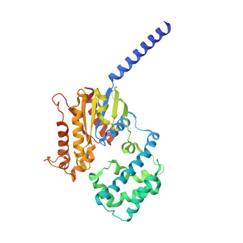
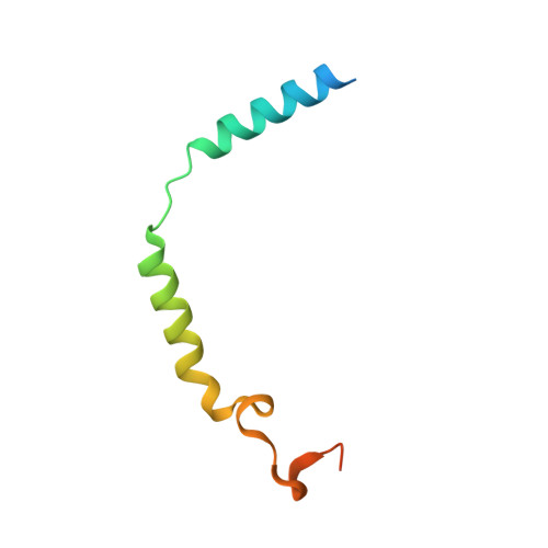
    G protein-coupled receptor-mediated heterotrimeric G protein activation is a major mode of signal transduction in the cell. Previously, we and other groups reported that the α5 helix of Gαi1, especially the hydrophobic interactions in this region, plays a key role during nucleotide release and G protein activation. To further investigate the effect of this hydrophobic core, we disrupted it in Gαi1 by inserting 4 alanine amino acids into the α5 helix between residues Gln(333) and Phe(334) (Ins4A). This extends the length of the α5 helix without disturbing the β6-α5 loop interactions. This mutant has high basal nucleotide exchange activity yet no receptor-mediated activation of nucleotide exchange. By using structural approaches, we show that this mutant loses critical hydrophobic interactions, leading to significant rearrangements of side chain residues His(57), Phe(189), Phe(191), and Phe(336); it also disturbs the rotation of the α5 helix and the π-π interaction between His(57) and Phe(189) In addition, the insertion mutant abolishes G protein release from the activated receptor after nucleotide binding. Our biochemical and computational data indicate that the interactions between α5, α1, and β2-β3 are not only vital for GDP release during G protein activation, but they are also necessary for proper GTP binding (or GDP rebinding). Thus, our studies suggest that this hydrophobic interface is critical for accurate rearrangement of the α5 helix for G protein release from the receptor after GTP binding.


  • Organizational Affiliation

    From the Departments of Pharmacology.


Macromolecules
Find similar proteins by:  (by identity cutoff)  |  3D Structure
Entity ID: 1
MoleculeChains Sequence LengthOrganismDetailsImage
Guanine nucleotide-binding protein G(i) subunit alpha-1358Rattus norvegicusMutation(s): 0 
Gene Names: Gnai1Gnai-1
UniProt
Find proteins for P10824 (Rattus norvegicus)
Explore P10824 
Go to UniProtKB:  P10824
Entity Groups  
Sequence Clusters30% Identity50% Identity70% Identity90% Identity95% Identity100% Identity
UniProt GroupP10824
Sequence Annotations
Expand
  • Reference Sequence
Find similar proteins by:  (by identity cutoff)  |  3D Structure
Entity ID: 2
MoleculeChains Sequence LengthOrganismDetailsImage
Guanine nucleotide-binding protein G(I)/G(S)/G(T) subunit beta-1340Bos taurusMutation(s): 0 
Gene Names: GNB1
UniProt
Find proteins for P62871 (Bos taurus)
Explore P62871 
Go to UniProtKB:  P62871
Entity Groups  
Sequence Clusters30% Identity50% Identity70% Identity90% Identity95% Identity100% Identity
UniProt GroupP62871
Sequence Annotations
Expand
  • Reference Sequence
Find similar proteins by:  (by identity cutoff)  |  3D Structure
Entity ID: 3
MoleculeChains Sequence LengthOrganismDetailsImage
Guanine nucleotide-binding protein G(T) subunit gamma-T1C [auth G]74Bos taurusMutation(s): 0 
Gene Names: GNGT1
UniProt
Find proteins for P02698 (Bos taurus)
Explore P02698 
Go to UniProtKB:  P02698
Entity Groups  
Sequence Clusters30% Identity50% Identity70% Identity90% Identity95% Identity100% Identity
UniProt GroupP02698
Sequence Annotations
Expand
  • Reference Sequence
Small Molecules
Ligands 1 Unique
IDChains Name / Formula / InChI Key2D Diagram3D Interactions
GDP
Query on GDP

Download Ideal Coordinates CCD File 
D [auth A]GUANOSINE-5'-DIPHOSPHATE
C10 H15 N5 O11 P2
QGWNDRXFNXRZMB-UUOKFMHZSA-N
Experimental Data & Validation

Experimental Data

  • Method: X-RAY DIFFRACTION
  • Resolution: 1.90 Å
  • R-Value Free: 0.208 
  • R-Value Work: 0.182 
  • R-Value Observed: 0.185 
  • Space Group: P 43
Unit Cell:
Length ( Å )Angle ( ˚ )
a = 84.648α = 90
b = 84.648β = 90
c = 130.091γ = 90
Software Package:
Software NamePurpose
PHENIXrefinement
HKL-2000data reduction
HKL-2000data scaling
PHENIXphasing

Structure Validation

View Full Validation Report


Ligand Structure Quality Assessment 


Entry History & Funding Information

Deposition Data


Funding OrganizationLocationGrant Number
National Institutes of Health/National Eye Institute (NIH/NEI)United StatesRO1 EY006062
National Institutes of Health/National Institute of General Medical Sciences (NIH/NIGMS)United StatesRO1 GM120569
National Institutes of Health/National Institute of General Medical Sciences (NIH/NIGMS)United StatesRO1 GM080403
National Institutes of Health/National Center for Research Resources (NIH/NCRR)United StatesS10 RR026915

Revision History  (Full details and data files)

  • Version 1.0: 2016-08-03
    Type: Initial release
  • Version 1.1: 2016-08-10
    Changes: Database references
  • Version 1.2: 2016-09-21
    Changes: Database references
  • Version 1.3: 2017-09-13
    Changes: Author supporting evidence, Database references, Derived calculations
  • Version 1.4: 2019-12-04
    Changes: Author supporting evidence
  • Version 1.5: 2023-09-27
    Changes: Data collection, Database references, Refinement description