5KAS

Murine acid sphingomyelinase-like phosphodiesterase 3b (SMPDL3B) with phosphocholine


Experimental Data Snapshot

  • Method: X-RAY DIFFRACTION
  • Resolution: 1.62 Å
  • R-Value Free: 0.193 
  • R-Value Work: 0.169 
  • R-Value Observed: 0.170 

wwPDB Validation   3D Report Full Report


Ligand Structure Quality Assessment 


This is version 2.0 of the entry. See complete history


Literature

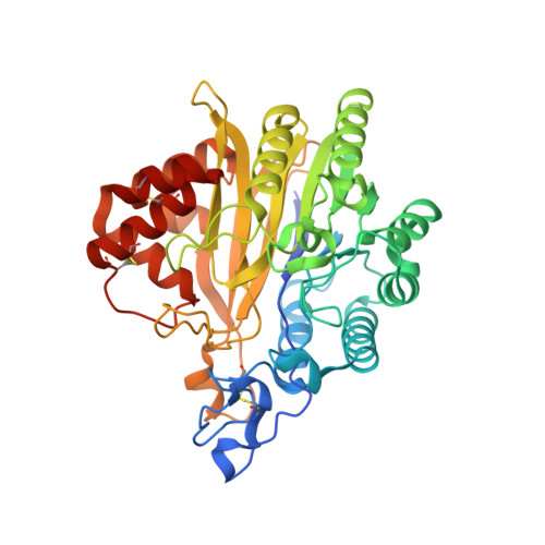
Crystal Structure of the Acid Sphingomyelinase-like Phosphodiesterase SMPDL3B Provides Insights into Determinants of Substrate Specificity.

Gorelik, A.Heinz, L.X.Illes, K.Superti-Furga, G.Nagar, B.

(2016) J Biol Chem 291: 24054-24064

  • DOI: https://doi.org/10.1074/jbc.M116.755801
  • Primary Citation of Related Structures:  
    5KAR, 5KAS

  • PubMed Abstract: 

    The enzyme acid sphingomyelinase-like phosphodiesterase 3B (SMPDL3B) was shown to act as a negative regulator of innate immune signaling, affecting cellular lipid composition and membrane fluidity. Furthermore, several reports identified this enzyme as an off target of the therapeutic antibody rituximab, with implications in kidney disorders. However, structural information for this protein is lacking. Here we present the high resolution crystal structure of murine SMPDL3B, which reveals a substrate binding site strikingly different from its paralogs. The active site is located in a narrow boot-shaped cavity. We identify a unique loop near the active site that appears to impose size constraints on incoming substrates. A structure in complex with phosphocholine indicates that the protein recognizes this head group via an aromatic box, a typical choline-binding motif. Although a potential substrate for SMPDL3B is sphingomyelin, we identify other possible substrates such as CDP-choline, ATP, and ADP. Functional experiments employing structure-guided mutagenesis in macrophages highlight amino acid residues potentially involved in recognition of endogenous substrates. Our study is an important step toward elucidating the specific function of this poorly characterized enzyme.


  • Organizational Affiliation

    From the Department of Biochemistry and Groupe de Recherche Axé sur la Structure des Protéines, McGill University, Montreal, Quebec H3G 0B1, Canada.


Macromolecules
Find similar proteins by:  (by identity cutoff)  |  3D Structure
Entity ID: 1
MoleculeChains Sequence LengthOrganismDetailsImage
Acid sphingomyelinase-like phosphodiesterase 3b427Mus musculusMutation(s): 0 
Gene Names: Smpdl3bAsml3b
EC: 3.1.4
UniProt
Find proteins for P58242 (Mus musculus)
Explore P58242 
Go to UniProtKB:  P58242
Entity Groups  
Sequence Clusters30% Identity50% Identity70% Identity90% Identity95% Identity100% Identity
UniProt GroupP58242
Sequence Annotations
Expand
  • Reference Sequence
Oligosaccharides

Help

Entity ID: 2
MoleculeChains Length2D Diagram Glycosylation3D Interactions
2-acetamido-2-deoxy-beta-D-glucopyranose-(1-4)-2-acetamido-2-deoxy-beta-D-glucopyranose
B
2N-Glycosylation
Glycosylation Resources
GlyTouCan:  G42666HT
GlyCosmos:  G42666HT
GlyGen:  G42666HT
Entity ID: 3
MoleculeChains Length2D Diagram Glycosylation3D Interactions
alpha-D-mannopyranose-(1-3)-beta-D-mannopyranose-(1-4)-2-acetamido-2-deoxy-beta-D-glucopyranose-(1-4)-2-acetamido-2-deoxy-beta-D-glucopyranose
C
4N-Glycosylation
Glycosylation Resources
GlyTouCan:  G81315DD
GlyCosmos:  G81315DD
GlyGen:  G81315DD
Entity ID: 4
MoleculeChains Length2D Diagram Glycosylation3D Interactions
2-acetamido-2-deoxy-beta-D-glucopyranose-(1-4)-[alpha-L-fucopyranose-(1-6)]2-acetamido-2-deoxy-beta-D-glucopyranose
D
3N-Glycosylation
Glycosylation Resources
GlyTouCan:  G21290RB
GlyCosmos:  G21290RB
GlyGen:  G21290RB
Experimental Data & Validation

Experimental Data

  • Method: X-RAY DIFFRACTION
  • Resolution: 1.62 Å
  • R-Value Free: 0.193 
  • R-Value Work: 0.169 
  • R-Value Observed: 0.170 
  • Space Group: P 1 2 1
Unit Cell:
Length ( Å )Angle ( ˚ )
a = 50.892α = 90
b = 47.031β = 98.07
c = 93.861γ = 90
Software Package:
Software NamePurpose
PHENIXrefinement
HKL-2000data reduction
HKL-2000data scaling
PHENIXphasing

Structure Validation

View Full Validation Report


Ligand Structure Quality Assessment 


Entry History & Funding Information

Deposition Data


Funding OrganizationLocationGrant Number
Canadian Institutes of Health Research (CIHR)CanadaMOP-133535

Revision History  (Full details and data files)

  • Version 1.0: 2016-10-05
    Type: Initial release
  • Version 1.1: 2016-10-12
    Changes: Database references
  • Version 1.2: 2016-11-23
    Changes: Database references
  • Version 1.3: 2020-01-08
    Changes: Author supporting evidence, Data collection, Database references
  • Version 2.0: 2020-07-29
    Type: Remediation
    Reason: Carbohydrate remediation
    Changes: Advisory, Atomic model, Data collection, Derived calculations, Structure summary