


Crystal Structure of an 8-amino-7-oxononanoate Synthase from Burkholderia xenovorans
Dranow, D.M., Conrady, D.G., Lorimer, D., Edwards, T.E.To be published.
Experimental Data Snapshot
wwPDB Validation 3D Report Full Report

Entity ID: 1 | |||||
|---|---|---|---|---|---|
| Molecule | Chains | Sequence Length | Organism | Details | Image |
| 8-amino-7-oxononanoate synthase | 402 | Paraburkholderia xenovorans LB400 | Mutation(s): 1 Gene Names: bioF, Bxeno_A0198, Bxe_A4264 EC: 2.3.1.47 | 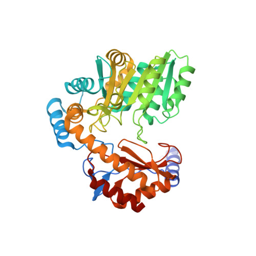 | |
UniProt | |||||
Find proteins for Q146K3 (Paraburkholderia xenovorans (strain LB400)) Explore Q146K3 Go to UniProtKB: Q146K3 | |||||
Entity Groups | |||||
| Sequence Clusters | 30% Identity50% Identity70% Identity90% Identity95% Identity100% Identity | ||||
| UniProt Group | Q146K3 | ||||
Sequence AnnotationsExpand | |||||
| |||||
| Length ( Å ) | Angle ( ˚ ) |
|---|---|
| a = 171.87 | α = 90 |
| b = 63.2 | β = 90 |
| c = 65.19 | γ = 90 |
| Software Name | Purpose |
|---|---|
| XSCALE | data scaling |
| PHENIX | refinement |
| PDB_EXTRACT | data extraction |
| XDS | data reduction |
| MOLREP | phasing |
| ARP | model building |
| Coot | model building |
RCSB PDB (citation) is hosted by
RCSB PDB is a member of the

