5J58

Trypanosoma brucei methionyl-tRNA synthetase in complex with inhibitor (Chem 1856)


Experimental Data Snapshot

  • Method: X-RAY DIFFRACTION
  • Resolution: 2.80 Å
  • R-Value Free: 0.230 
  • R-Value Work: 0.202 
  • R-Value Observed: 0.203 

wwPDB Validation   3D Report Full Report


Ligand Structure Quality Assessment 


This is version 1.1 of the entry. See complete history


Literature

Structure-guided design of novel Trypanosoma brucei Methionyl-tRNA synthetase inhibitors.

Huang, W.Zhang, Z.Barros-Alvarez, X.Koh, C.Y.Ranade, R.M.Gillespie, J.R.Creason, S.A.Shibata, S.Verlinde, C.L.Hol, W.G.Buckner, F.S.Fan, E.

(2016) Eur J Med Chem 124: 1081-1092

  • DOI: https://doi.org/10.1016/j.ejmech.2016.10.024
  • Primary Citation of Related Structures:  
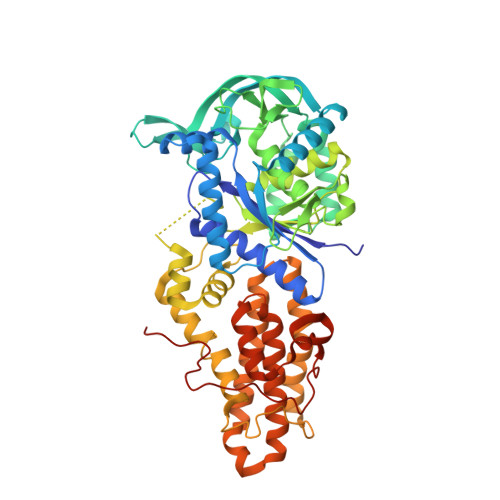
    5J58, 5J59, 5J5A

  • PubMed Abstract: 

    A screening hit 1 against Trypanosoma brucei methionyl-tRNA synthetase was optimized using a structure-guided approach. The optimization led to the identification of two novel series of potent inhibitors, the cyclic linker and linear linker series. Compounds of both series were potent in a T. brucei growth inhibition assay while showing low toxicity to mammalian cells. The best compound of each series, 16 and 31, exhibited EC 50 s of 39 and 22 nM, respectively. Compounds 16 and 31 also exhibited promising PK properties after oral dosing in mice. Moreover, compound 31 had moderately good brain permeability, with a brain/plasma ratio of 0.27 at 60 min after IP injection. This study provides new lead compounds for arriving at new treatments of human African trypanosomiasis (HAT).


  • Organizational Affiliation

    Department of Biochemistry, University of Washington, Seattle, WA 98195, United States.


Macromolecules
Find similar proteins by:  (by identity cutoff)  |  3D Structure
Entity ID: 1
MoleculeChains Sequence LengthOrganismDetailsImage
Methionyl-tRNA synthetase, putative
A, B
542Trypanosoma brucei brucei TREU927Mutation(s): 6 
Gene Names: Tb10.70.6470
EC: 6.1.1.10
UniProt
Find proteins for Q38C91 (Trypanosoma brucei brucei (strain 927/4 GUTat10.1))
Explore Q38C91 
Go to UniProtKB:  Q38C91
Entity Groups  
Sequence Clusters30% Identity50% Identity70% Identity90% Identity95% Identity100% Identity
UniProt GroupQ38C91
Sequence Annotations
Expand
  • Reference Sequence
Small Molecules
Ligands 4 Unique
IDChains Name / Formula / InChI Key2D Diagram3D Interactions
N56
Query on N56

Download Ideal Coordinates CCD File 
J [auth B](3S)-1-(5-chloro-1H-imidazo[4,5-b]pyridin-2-yl)-N-[(3,5-dichlorophenyl)methyl]piperidin-3-amine
C18 H18 Cl3 N5
ALKJREGOUJGYEK-AWEZNQCLSA-N
MET
Query on MET

Download Ideal Coordinates CCD File 
C [auth A]METHIONINE
C5 H11 N O2 S
FFEARJCKVFRZRR-BYPYZUCNSA-N
GOL
Query on GOL

Download Ideal Coordinates CCD File 
D [auth A],
E [auth A],
H [auth B]
GLYCEROL
C3 H8 O3
PEDCQBHIVMGVHV-UHFFFAOYSA-N
DMS
Query on DMS

Download Ideal Coordinates CCD File 
F [auth A],
G [auth A],
I [auth B]
DIMETHYL SULFOXIDE
C2 H6 O S
IAZDPXIOMUYVGZ-UHFFFAOYSA-N
Experimental Data & Validation

Experimental Data

  • Method: X-RAY DIFFRACTION
  • Resolution: 2.80 Å
  • R-Value Free: 0.230 
  • R-Value Work: 0.202 
  • R-Value Observed: 0.203 
  • Space Group: P 21 21 21
Unit Cell:
Length ( Å )Angle ( ˚ )
a = 86.97α = 90
b = 106.03β = 90
c = 207.31γ = 90
Software Package:
Software NamePurpose
REFMACrefinement
PHASERphasing
Aimlessdata scaling
HKL-2000data reduction

Structure Validation

View Full Validation Report


Ligand Structure Quality Assessment 


Entry History 

Deposition Data

Revision History  (Full details and data files)

  • Version 1.0: 2017-01-25
    Type: Initial release
  • Version 1.1: 2024-03-06
    Changes: Data collection, Database references, Refinement description