


Crystal Structure of Cobyrinic Acid a,c-diamide synthase from Mycobacterium smegmatis with bound ATP analog and Magnesium
Davies, D.R., Dranow, D.M., Abendroth, J.A., SSGCIDTo be published.
Experimental Data Snapshot
Entity ID: 1 | |||||
|---|---|---|---|---|---|
| Molecule | Chains | Sequence Length | Organism | Details | Image |
| Cobyrinic Acid a,c-diamide synthase | 273 | Mycolicibacterium smegmatis MC2 155 | Mutation(s): 0 Gene Names: MSMEG_1927 | 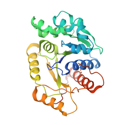 | |
UniProt | |||||
Find proteins for A0QTQ5 (Mycolicibacterium smegmatis (strain ATCC 700084 / mc(2)155)) Explore A0QTQ5 Go to UniProtKB: A0QTQ5 | |||||
Entity Groups | |||||
| Sequence Clusters | 30% Identity50% Identity70% Identity90% Identity95% Identity100% Identity | ||||
| UniProt Group | A0QTQ5 | ||||
Sequence AnnotationsExpand | |||||
| |||||
| Ligands 2 Unique | |||||
|---|---|---|---|---|---|
| ID | Chains | Name / Formula / InChI Key | 2D Diagram | 3D Interactions | |
| ANP Query on ANP | C [auth A], E [auth B] | PHOSPHOAMINOPHOSPHONIC ACID-ADENYLATE ESTER C10 H17 N6 O12 P3 PVKSNHVPLWYQGJ-KQYNXXCUSA-N |  | ||
| MG Query on MG | D [auth A], F [auth B] | MAGNESIUM ION Mg JLVVSXFLKOJNIY-UHFFFAOYSA-N |  | ||
| Length ( Å ) | Angle ( ˚ ) |
|---|---|
| a = 88.4 | α = 90 |
| b = 88.4 | β = 90 |
| c = 120.3 | γ = 90 |
| Software Name | Purpose |
|---|---|
| PHENIX | refinement |
| XDS | data reduction |
| XSCALE | data scaling |
| PHASER | phasing |
RCSB PDB (citation) is hosted by
RCSB PDB is a member of the

