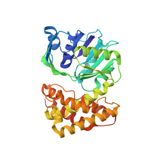
Crystal structure of ketopantoate reductase from Thermococcus kodakarensis complexed with NADP+
Aikawa, Y., Nishitani, Y., Tomita, H., Atomi, H., Miki, K.(2016) Acta Crystallogr F Struct Biol Commun 72: 369-375
- PubMed: 27139828
- DOI: https://doi.org/10.1107/S2053230X16005033
- Primary Citation of Related Structures:
5HWS - PubMed Abstract:
Coenzyme A (CoA) plays pivotal roles in a variety of metabolic pathways in all organisms. The biosynthetic pathway of CoA is strictly regulated by feedback inhibition. In the hyperthermophilic archaeon Thermococcus kodakarensis, ketopantoate reductase (KPR), which catalyzes the NAD(P)H-dependent reduction of 2-oxopantoate, is a target of feedback inhibition by CoA. The crystal structure of KPR from T. kodakarensis (Tk-KPR) complexed with CoA and 2-oxopantoate has previously been reported. The structure provided an explanation for the competitive inhibition mechanism. Here, further biochemical analyses of Tk-KPR and the crystal structure of Tk-KPR in complex with NADP(+) are reported. A mutational analysis implies that the residues in the binding pocket cooperatively contribute to the recognition of CoA. The structure reveals the same dimer architecture as the Tk-KPR-CoA-2-oxopantoate complex. Moreover, the positions of the residues involved in the dimer interaction are not changed by the binding of CoA and 2-oxopantoate, suggesting individual conformational changes of Tk-KPR monomers.
Organizational Affiliation:
Department of Chemistry, Graduate School of Science, Kyoto University, Sakyo-ku, Kyoto 606-8502, Japan.