5HP3

Human Adenosine Deaminase Acting on dsRNA (ADAR2) bound to dsRNA sequence derived from S. cerevisiae BDF2 gene with AC mismatch at reaction site


Experimental Data Snapshot

  • Method: X-RAY DIFFRACTION
  • Resolution: 3.09 Å
  • R-Value Free: 0.238 
  • R-Value Work: 0.163 
  • R-Value Observed: 0.167 

wwPDB Validation   3D Report Full Report


Ligand Structure Quality Assessment 


This is version 1.7 of the entry. See complete history


Literature

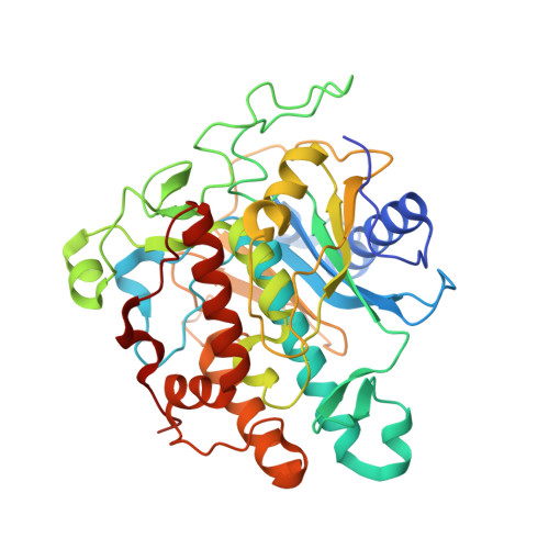
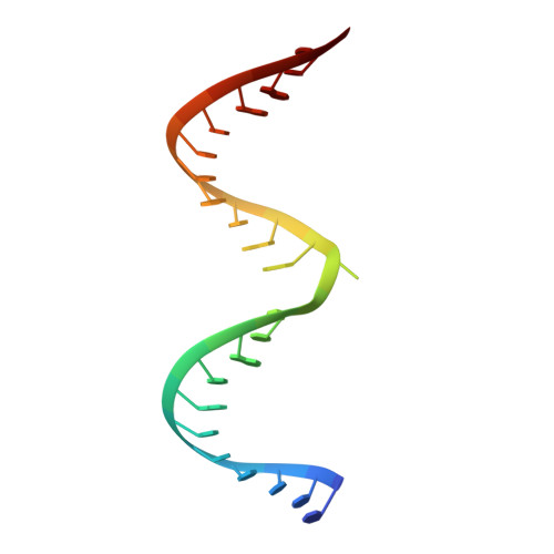
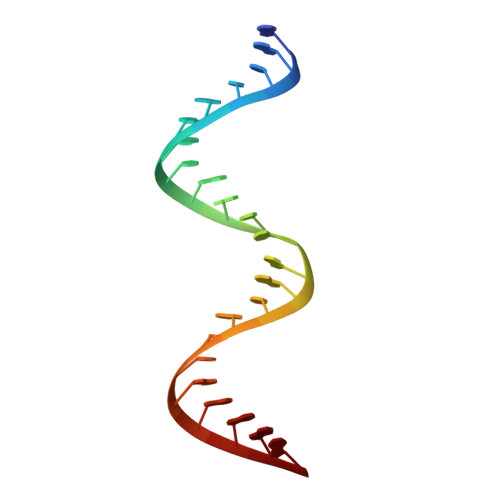
Structures of human ADAR2 bound to dsRNA reveal base-flipping mechanism and basis for site selectivity.

Matthews, M.M.Thomas, J.M.Zheng, Y.Tran, K.Phelps, K.J.Scott, A.I.Havel, J.Fisher, A.J.Beal, P.A.

(2016) Nat Struct Mol Biol 23: 426-433

  • DOI: https://doi.org/10.1038/nsmb.3203
  • Primary Citation of Related Structures:  
    5ED1, 5ED2, 5HP2, 5HP3

  • PubMed Abstract: 

    Adenosine deaminases acting on RNA (ADARs) are editing enzymes that convert adenosine to inosine in duplex RNA, a modification reaction with wide-ranging consequences in RNA function. Understanding of the ADAR reaction mechanism, the origin of editing-site selectivity, and the effect of mutations is limited by the lack of high-resolution structural data for complexes of ADARs bound to substrate RNAs. Here we describe four crystal structures of the human ADAR2 deaminase domain bound to RNA duplexes bearing a mimic of the deamination reaction intermediate. These structures, together with structure-guided mutagenesis and RNA-modification experiments, explain the basis of the ADAR deaminase domain's dsRNA specificity, its base-flipping mechanism, and its nearest-neighbor preferences. In addition, we identified an ADAR2-specific RNA-binding loop near the enzyme active site, thus rationalizing differences in selectivity observed between different ADARs. Finally, our results provide a structural framework for understanding the effects of ADAR mutations associated with human disease.


  • Organizational Affiliation

    Department of Chemistry, University of California, Davis, Davis, California, USA.


Macromolecules

Find similar proteins by:  (by identity cutoff)  |  3D Structure
Entity ID: 1
MoleculeChains Sequence LengthOrganismDetailsImage
Double-stranded RNA-specific editase 1
A, D
403Homo sapiensMutation(s): 0 
Gene Names: ADARB1ADAR2DRADA2RED1
EC: 3.5.4.37
UniProt & NIH Common Fund Data Resources
Find proteins for P78563 (Homo sapiens)
Explore P78563 
Go to UniProtKB:  P78563
PHAROS:  P78563
GTEx:  ENSG00000197381 
Entity Groups  
Sequence Clusters30% Identity50% Identity70% Identity90% Identity95% Identity100% Identity
UniProt GroupP78563
Sequence Annotations
Expand
  • Reference Sequence

Find similar nucleic acids by:  Sequence   |   3D Structure  

Entity ID: 2
MoleculeChains LengthOrganismImage
RNA (5'-R(*UP*UP*CP*CP*CP*CP*AP*CP*AP*UP*UP*(8AZ)P*GP*AP*CP*GP*UP*UP*CP*AP*GP*UP*C)-3')23Saccharomyces cerevisiae
Sequence Annotations
Expand
  • Reference Sequence

Find similar nucleic acids by:  Sequence   |   3D Structure  

Entity ID: 3
MoleculeChains LengthOrganismImage
RNA (5'-R(*GP*AP*CP*UP*GP*AP*AP*CP*GP*AP*CP*CP*AP*AP*UP*GP*UP*GP*GP*GP*GP*AP*A)-3')23Saccharomyces cerevisiae
Sequence Annotations
Expand
  • Reference Sequence
Experimental Data & Validation

Experimental Data

  • Method: X-RAY DIFFRACTION
  • Resolution: 3.09 Å
  • R-Value Free: 0.238 
  • R-Value Work: 0.163 
  • R-Value Observed: 0.167 
  • Space Group: P 21 21 21
Unit Cell:
Length ( Å )Angle ( ˚ )
a = 81.51α = 90
b = 107.21β = 90
c = 120.62γ = 90
Software Package:
Software NamePurpose
PHENIXrefinement
PHENIXmodel building
XSCALEdata scaling
XDSdata reduction
PHASERphasing

Structure Validation

View Full Validation Report


Ligand Structure Quality Assessment 


Entry History & Funding Information

Deposition Data


Funding OrganizationLocationGrant Number
National Institutes of Health/National Institute of General Medical Sciences (NIH/NIGMS)United StatesR01GM061115

Revision History  (Full details and data files)

  • Version 1.0: 2016-04-13
    Type: Initial release
  • Version 1.1: 2016-04-27
    Changes: Database references
  • Version 1.2: 2016-05-18
    Changes: Database references
  • Version 1.3: 2017-09-13
    Changes: Author supporting evidence, Derived calculations
  • Version 1.4: 2019-12-25
    Changes: Author supporting evidence
  • Version 1.5: 2020-10-14
    Changes: Derived calculations, Structure summary
  • Version 1.6: 2021-02-10
    Changes: Derived calculations
  • Version 1.7: 2023-09-27
    Changes: Data collection, Database references, Refinement description