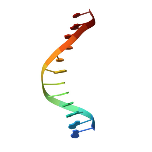
5HNK

Crystal structure of T5Fen in complex intact substrate and metal ions.


Experimental Data Snapshot

  • Method: X-RAY DIFFRACTION
  • Resolution: 2.22 Å
  • R-Value Free: 0.234 
  • R-Value Work: 0.183 
  • R-Value Observed: 0.185 

wwPDB Validation   3D Report Full Report


This is version 1.4 of the entry. See complete history


Literature

Direct observation of DNA threading in flap endonuclease complexes.

AlMalki, F.A.Flemming, C.S.Zhang, J.Feng, M.Sedelnikova, S.E.Ceska, T.Rafferty, J.B.Sayers, J.R.Artymiuk, P.J.

(2016) Nat Struct Mol Biol 23: 640-646

  • DOI: https://doi.org/10.1038/nsmb.3241
  • Primary Citation of Related Structures:  
    5HML, 5HMM, 5HNK, 5HP4

  • PubMed Abstract: 

    Maintenance of genome integrity requires that branched nucleic acid molecules be accurately processed to produce double-helical DNA. Flap endonucleases are essential enzymes that trim such branched molecules generated by Okazaki-fragment synthesis during replication. Here, we report crystal structures of bacteriophage T5 flap endonuclease in complexes with intact DNA substrates and products, at resolutions of 1.9-2.2 Å. They reveal single-stranded DNA threading through a hole in the enzyme, which is enclosed by an inverted V-shaped helical arch straddling the active site. Residues lining the hole induce an unusual barb-like conformation in the DNA substrate, thereby juxtaposing the scissile phosphate and essential catalytic metal ions. A series of complexes and biochemical analyses show how the substrate's single-stranded branch approaches, threads through and finally emerges on the far side of the enzyme. Our studies suggest that substrate recognition involves an unusual 'fly-casting, thread, bend and barb' mechanism.


  • Organizational Affiliation

    Department of Molecular Biology and Biotechnology, University of Sheffield, Sheffield, UK.


Macromolecules

Find similar proteins by:  (by identity cutoff)  |  3D Structure
Entity ID: 2
MoleculeChains Sequence LengthOrganismDetailsImage
ExodeoxyribonucleaseC [auth A],
D [auth B]
272Tequintavirus T5Mutation(s): 1 
Gene Names: D15
EC: 3.1.11.3
UniProt
Find proteins for P06229 (Escherichia phage T5)
Explore P06229 
Go to UniProtKB:  P06229
Entity Groups  
Sequence Clusters30% Identity50% Identity70% Identity90% Identity95% Identity100% Identity
UniProt GroupP06229
Sequence Annotations
Expand
  • Reference Sequence

Find similar nucleic acids by:  Sequence   |   3D Structure  

Entity ID: 1
MoleculeChains LengthOrganismImage
DNA (5'-D(*AP*AP*AP*AP*GP*CP*GP*TP*AP*CP*GP*C)-3')A [auth X],
B [auth Y]
12synthetic construct
Sequence Annotations
Expand
  • Reference Sequence
Experimental Data & Validation

Experimental Data

  • Method: X-RAY DIFFRACTION
  • Resolution: 2.22 Å
  • R-Value Free: 0.234 
  • R-Value Work: 0.183 
  • R-Value Observed: 0.185 
  • Space Group: P 21 21 21
Unit Cell:
Length ( Å )Angle ( ˚ )
a = 44.72α = 90
b = 109.94β = 90
c = 127.34γ = 90
Software Package:
Software NamePurpose
SCALAdata scaling
REFMACrefinement
PDB_EXTRACTdata extraction
xia2data reduction
PHASERphasing

Structure Validation

View Full Validation Report


Entry History 

Deposition Data

Revision History  (Full details and data files)

  • Version 1.0: 2016-06-01
    Type: Initial release
  • Version 1.1: 2016-06-08
    Changes: Database references
  • Version 1.2: 2016-06-22
    Changes: Database references
  • Version 1.3: 2016-07-20
    Changes: Database references
  • Version 1.4: 2024-01-10
    Changes: Data collection, Database references, Derived calculations, Refinement description