


Crystal structure of a hypothetical protein from Synechocystis
Dong, L.-L., Wu, D., Gao, X.-Q., Zhou, M., Liu, Z.-J., Zhao, K.-H.To be published.
Experimental Data Snapshot
wwPDB Validation 3D Report Full Report

Entity ID: 1 | |||||
|---|---|---|---|---|---|
| Molecule | Chains | Sequence Length | Organism | Details | Image |
| Slr0280 protein | 610 | Synechocystis sp. PCC 6803 substr. Kazusa | Mutation(s): 0 Gene Names: slr0280 | 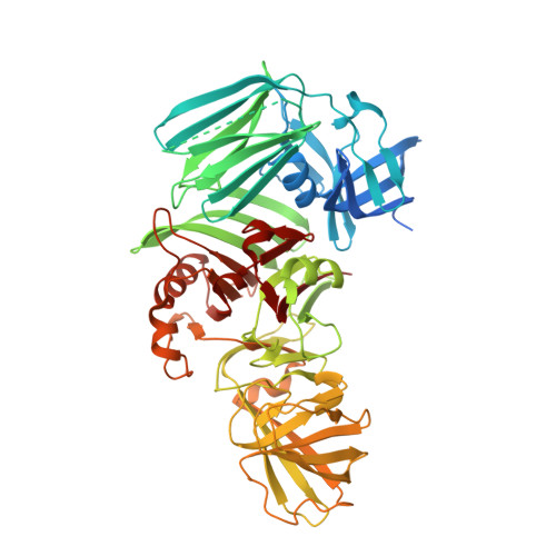 | |
UniProt | |||||
Find proteins for P74396 (Synechocystis sp. (strain PCC 6803 / Kazusa)) Explore P74396 Go to UniProtKB: P74396 | |||||
Entity Groups | |||||
| Sequence Clusters | 30% Identity50% Identity70% Identity90% Identity95% Identity100% Identity | ||||
| UniProt Group | P74396 | ||||
Sequence AnnotationsExpand | |||||
| |||||
| Length ( Å ) | Angle ( ˚ ) |
|---|---|
| a = 66.152 | α = 90 |
| b = 83.924 | β = 90 |
| c = 96.069 | γ = 90 |
| Software Name | Purpose |
|---|---|
| PHENIX | refinement |
| HKL-2000 | data reduction |
| HKL-2000 | data scaling |
| PHENIX | phasing |
RCSB PDB (citation) is hosted by
RCSB PDB is a member of the

