5GTO

Human PPARgamma ligand binding dmain complexed with S35


Experimental Data Snapshot

  • Method: X-RAY DIFFRACTION
  • Resolution: 2.10 Å
  • R-Value Free: 0.242 
  • R-Value Work: 0.211 
  • R-Value Observed: 0.212 

wwPDB Validation   3D Report Full Report


Ligand Structure Quality Assessment 


This is version 1.2 of the entry. See complete history


Literature

Structural basis for differential activities of enantiomeric PPAR gamma agonists: Binding of S35 to the alternate site.

Jang, J.Y.Koh, M.Bae, H.An, D.R.Im, H.N.Kim, H.S.Yoon, J.Y.Yoon, H.J.Han, B.W.Park, S.B.Suh, S.W.

(2017) Biochim Biophys Acta 1865: 674-681

  • DOI: https://doi.org/10.1016/j.bbapap.2017.03.008
  • Primary Citation of Related Structures:  
    5GTN, 5GTO, 5GTP

  • PubMed Abstract: 

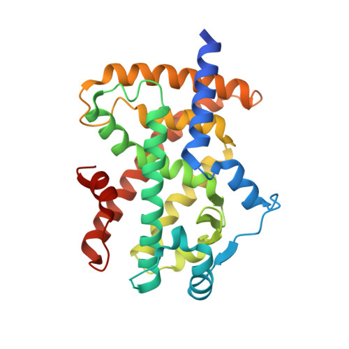
    Peroxisome proliferator-activated receptor γ (PPARγ) is a member of the nuclear receptor superfamily. It functions as a ligand-activated transcription factor and plays important roles in the regulation of adipocyte differentiation, type 2 diabetes mellitus, and inflammation. Many PPARγ agonists bind to the canonical ligand-binding pocket near the activation function-2 (AF-2) helix (i.e., helix H12) of the ligand-binding domain (LBD). More recently, an alternate ligand-binding site was identified in PPARγ LBD; it is located beside the Ω loop between the helices H2' and H3. We reported previously that the chirality of two optimized enantiomeric PPARγ ligands (S35 and R35) differentiates their PPARγ transcriptional activity, binding affinity, and inhibitory activity toward Cdk5 (cyclin-dependent kinase 5)-mediated phosphorylation of PPARγ at Ser245 (in PPARγ1 numbering; Ser273 in PPARγ2 numbering). S35 is a PPARγ phosphorylation inhibitor with promising glucose uptake potential, whereas R35 behaves as a potent conventional PPARγ agonist. To provide a structural basis for understanding the differential activities of these enantiomeric ligands, we have determined crystal structures of the PPARγ LBD in complex with either S35 or R35. S35 and R35 bind to the PPARγ LBD in significantly different manners. The partial agonist S35 occupies the alternate site near the Ω loop, whereas the full agonist R35 binds entirely to the canonical LBP. Alternate site binding of S35 affects the PPARγ transactivation and the inhibitory effect on PPARγ Ser245 phosphorylation. This study provides a useful platform for the development of a new generation of PPARγ ligands as anti-diabetic drug candidates.


  • Organizational Affiliation

    Department of Chemistry, College of Natural Sciences, Seoul National University, Seoul 08826, Republic of Korea; Research Institute of Pharmaceutical Sciences, College of Pharmacy, Seoul National University, Seoul 08826, Republic of Korea.


Macromolecules
Find similar proteins by:  (by identity cutoff)  |  3D Structure
Entity ID: 1
MoleculeChains Sequence LengthOrganismDetailsImage
Peroxisome proliferator-activated receptor gamma283Homo sapiensMutation(s): 0 
Gene Names: PPARGNR1C3
UniProt & NIH Common Fund Data Resources
Find proteins for P37231 (Homo sapiens)
Explore P37231 
Go to UniProtKB:  P37231
PHAROS:  P37231
GTEx:  ENSG00000132170 
Entity Groups  
Sequence Clusters30% Identity50% Identity70% Identity90% Identity95% Identity100% Identity
UniProt GroupP37231
Sequence Annotations
Expand
  • Reference Sequence

Find similar proteins by:  Sequence   |   3D Structure  

Entity ID: 2
MoleculeChains Sequence LengthOrganismDetailsImage
Nuclear receptor coactivator 116Homo sapiensMutation(s): 0 
UniProt & NIH Common Fund Data Resources
Find proteins for Q15788 (Homo sapiens)
Explore Q15788 
Go to UniProtKB:  Q15788
PHAROS:  Q15788
GTEx:  ENSG00000084676 
Entity Groups  
Sequence Clusters30% Identity50% Identity70% Identity90% Identity95% Identity100% Identity
UniProt GroupQ15788
Sequence Annotations
Expand
  • Reference Sequence
Small Molecules
Ligands 3 Unique
IDChains Name / Formula / InChI Key2D Diagram3D Interactions
T35
Query on T35

Download Ideal Coordinates CCD File 
C [auth A]2-[4-[5-[(1~{S})-1-[(3,5-dimethoxyphenyl)carbamoyl-(phenylmethyl)carbamoyl]oxypropyl]-1,2-oxazol-3-yl]phenoxy]-2-methyl-propanoic acid
C33 H35 N3 O9
WSCPDRVLOQMVLP-NDEPHWFRSA-N
MYR
Query on MYR

Download Ideal Coordinates CCD File 
D [auth A]MYRISTIC ACID
C14 H28 O2
TUNFSRHWOTWDNC-UHFFFAOYSA-N
GOL
Query on GOL

Download Ideal Coordinates CCD File 
E [auth A]GLYCEROL
C3 H8 O3
PEDCQBHIVMGVHV-UHFFFAOYSA-N
Binding Affinity Annotations 
IDSourceBinding Affinity
MYR BindingDB:  5GTO IC50: 2.10e+4 (nM) from 1 assay(s)
T35 BindingDB:  5GTO EC50: 183 (nM) from 1 assay(s)
Experimental Data & Validation

Experimental Data

  • Method: X-RAY DIFFRACTION
  • Resolution: 2.10 Å
  • R-Value Free: 0.242 
  • R-Value Work: 0.211 
  • R-Value Observed: 0.212 
  • Space Group: P 21 21 2
Unit Cell:
Length ( Å )Angle ( ˚ )
a = 131.823α = 90
b = 53.286β = 90
c = 53.74γ = 90
Software Package:
Software NamePurpose
REFMACrefinement
HKL-2000data reduction
HKL-2000data scaling
MOLREPphasing

Structure Validation

View Full Validation Report


Ligand Structure Quality Assessment 


Entry History 

Deposition Data

Revision History  (Full details and data files)

  • Version 1.0: 2017-07-05
    Type: Initial release
  • Version 1.1: 2017-08-30
    Changes: Data collection
  • Version 1.2: 2023-11-08
    Changes: Data collection, Database references, Refinement description