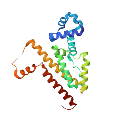
Modular Organisation of Inducer Recognition and Allostery in the Tetracycline Repressor
Werten, S., Schneider, J., Palm, G.J., Hinrichs, W.(2016) FEBS J 283: 2102
- PubMed: 27028290
- DOI: https://doi.org/10.1111/febs.13723
- Primary Citation of Related Structures:
5FKK, 5FKL, 5FKM, 5FKN, 5FKO - PubMed Abstract:
Induction of the tetracycline repressor (TetR) results from antibiotic-dependent changes in the relative positioning of the DNA-binding domains within the promoter-associated repressor dimer, but the key determinants of this allosteric effect remain poorly characterised. Intriguingly, previous mutational analyses of the tetracycline-interacting site revealed a lack of correlation between residual affinity and induction propensity, suggesting that some of the residues in contact with the antibiotic primarily act in ligand recognition and retention, whereas others are required to transmit the allosteric signal. Here, we provide a structural basis for these observations via crystallographic analysis of the point mutants N82A, H100A, T103A and E147A in complex with the inducer 5a,6-anhydrotetracycline. In conjunction with the available functional data, the four structures demonstrate that a trigger-like movement of the region between helices α6 and α7 towards and into the binding site plays a decisive role in the intramolecular communication process. In sharp contrast, residues lining the binding cavity proper have little or no influence on the allosteric mechanism as such. This nearly complete physical separation of ligand recognition and allostery will have allowed diverging TetR-like repressors to bind novel effectors while the existing induction mechanism remained intact. Consequently, the modularity described here may have been a key factor in the evolutionary success of the widespread and highly diversified repressor class.
Organizational Affiliation:
Department of Molecular Structural Biology, Institute for Biochemistry, University of Greifswald, Germany.


