5FJY

Crystal structure of mouse kinesin light chain 2 (residues 161-480)


Experimental Data Snapshot

  • Method: X-RAY DIFFRACTION
  • Resolution: 4.00 Å
  • R-Value Free: 0.261 
  • R-Value Work: 0.233 
  • R-Value Observed: 0.234 

wwPDB Validation   3D Report Full Report


This is version 1.3 of the entry. See complete history


Literature

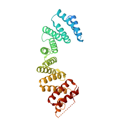
The Light Chains of Kinesin-1 are Autoinhibited.

Yip, Y.Y.Pernigo, S.Sanger, A.Xu, M.Parsons, M.Steiner, R.A.Dodding, M.P.

(2016) Proc Natl Acad Sci U S A 113: 2418

  • DOI: https://doi.org/10.1073/pnas.1520817113
  • Primary Citation of Related Structures:  
    5FJY

  • PubMed Abstract: 

    The light chains (KLCs) of the microtubule motor kinesin-1 bind cargoes and regulate its activity. Through their tetratricopeptide repeat domain (KLC(TPR)), they can recognize short linear peptide motifs found in many cargo proteins characterized by a central tryptophan flanked by aspartic/glutamic acid residues (W-acidic). Using a fluorescence resonance energy transfer biosensor in combination with X-ray crystallographic, biochemical, and biophysical approaches, we describe how an intramolecular interaction between the KLC2(TPR) domain and a conserved peptide motif within an unstructured region of the molecule, partly occludes the W-acidic binding site on the TPR domain. Cargo binding displaces this interaction, effecting a global conformational change in KLCs resulting in a more extended conformation. Thus, like the motor-bearing kinesin heavy chains, KLCs exist in a dynamic conformational state that is regulated by self-interaction and cargo binding. We propose a model by which, via this molecular switch, W-acidic cargo binding regulates the activity of the holoenzyme.


  • Organizational Affiliation

    Randall Division of Cell and Molecular Biophysics, King's College London, London SE1 1UL, United Kingdom.


Macromolecules
Find similar proteins by:  (by identity cutoff)  |  3D Structure
Entity ID: 1
MoleculeChains Sequence LengthOrganismDetailsImage
KINESIN LIGHT CHAIN 2
A, B, C
324Mus musculusMutation(s): 0 
UniProt & NIH Common Fund Data Resources
Find proteins for O88448 (Mus musculus)
Explore O88448 
Go to UniProtKB:  O88448
IMPC:  MGI:107953
Entity Groups  
Sequence Clusters30% Identity50% Identity70% Identity90% Identity95% Identity100% Identity
UniProt GroupO88448
Sequence Annotations
Expand
  • Reference Sequence

Find similar proteins by:  Sequence   |   3D Structure  

Entity ID: 2
MoleculeChains Sequence LengthOrganismDetailsImage
UNKNOWN PEPTIDE
D, E, F
5Mus musculusMutation(s): 0 
Sequence Annotations
Expand
  • Reference Sequence
Experimental Data & Validation

Experimental Data

  • Method: X-RAY DIFFRACTION
  • Resolution: 4.00 Å
  • R-Value Free: 0.261 
  • R-Value Work: 0.233 
  • R-Value Observed: 0.234 
  • Space Group: C 1 2 1
Unit Cell:
Length ( Å )Angle ( ˚ )
a = 148.7α = 90
b = 86.28β = 98.41
c = 111.74γ = 90
Software Package:
Software NamePurpose
BUSTERrefinement
XDSdata reduction
Aimlessdata scaling
PHASERphasing

Structure Validation

View Full Validation Report


Entry History 

Deposition Data

Revision History  (Full details and data files)

  • Version 1.0: 2016-02-24
    Type: Initial release
  • Version 1.1: 2016-03-02
    Changes: Database references
  • Version 1.2: 2016-03-16
    Changes: Database references
  • Version 1.3: 2024-01-10
    Changes: Data collection, Database references, Other, Refinement description