5FFD

CopM in the Ag-bound form (by co-crystallization)


Experimental Data Snapshot

  • Method: X-RAY DIFFRACTION
  • Resolution: 1.45 Å
  • R-Value Free: 0.193 
  • R-Value Work: 0.156 
  • R-Value Observed: 0.158 

wwPDB Validation   3D Report Full Report


This is version 1.2 of the entry. See complete history


Literature

Structural basis for copper/silver binding by the Synechocystis metallochaperone CopM.

Zhao, S.Wang, X.Niu, G.Dong, W.Wang, J.Fang, Y.Lin, Y.Liu, L.

(2016) Acta Crystallogr D Struct Biol 72: 997-1005

  • DOI: https://doi.org/10.1107/S2059798316011943
  • Primary Citation of Related Structures:  
    5FEJ, 5FFA, 5FFB, 5FFC, 5FFD, 5FFE

  • PubMed Abstract: 

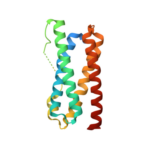
    Copper homeostasis integrates multiple processes from sensing to storage and efflux out of the cell. CopM is a cyanobacterial metallochaperone, the gene for which is located upstream of a two-component system for copper resistance, but the molecular basis for copper recognition by this four-helical bundle protein is unknown. Here, crystal structures of CopM in apo, copper-bound and silver-bound forms are reported. Monovalent copper/silver ions are buried within the bundle core; divalent copper ions are found on the surface of the bundle. The monovalent copper/silver-binding site is constituted by two consecutive histidines and is conserved in a previously functionally unknown protein family. The structural analyses show two conformational states and suggest that flexibility in the first α-helix is related to the metallochaperone function. These results also reveal functional diversity from a protein family with a simple four-helical fold.


  • Organizational Affiliation

    Key Laboratory of Photobiology, CAS Center for Excellence in Molecular Plant Sciences, Institute of Botany, Chinese Academy of Sciences, Beijing 100093, People's Republic of China.


Macromolecules
Find similar proteins by:  (by identity cutoff)  |  3D Structure
Entity ID: 1
MoleculeChains Sequence LengthOrganismDetailsImage
CopM172Synechocystis sp. PCC 6803Mutation(s): 0 
Gene Names: pcopM
UniProt
Find proteins for Q6YRW5 (Synechocystis sp. (strain PCC 6803 / Kazusa))
Explore Q6YRW5 
Go to UniProtKB:  Q6YRW5
Entity Groups  
Sequence Clusters30% Identity50% Identity70% Identity90% Identity95% Identity100% Identity
UniProt GroupQ6YRW5
Sequence Annotations
Expand
  • Reference Sequence
Experimental Data & Validation

Experimental Data

  • Method: X-RAY DIFFRACTION
  • Resolution: 1.45 Å
  • R-Value Free: 0.193 
  • R-Value Work: 0.156 
  • R-Value Observed: 0.158 
  • Space Group: P 21 21 2
Unit Cell:
Length ( Å )Angle ( ˚ )
a = 53.872α = 90
b = 85.959β = 90
c = 31.729γ = 90
Software Package:
Software NamePurpose
HKL-2000data collection
HKL-2000data scaling
PHASERphasing
PHENIXrefinement
PDB_EXTRACTdata extraction
HKL-2000data reduction
PHASERphasing

Structure Validation

View Full Validation Report


Entry History 

Deposition Data

Revision History  (Full details and data files)

  • Version 1.0: 2016-09-07
    Type: Initial release
  • Version 1.1: 2019-12-25
    Changes: Database references, Derived calculations
  • Version 1.2: 2023-11-08
    Changes: Data collection, Database references, Refinement description