5F84

Crystal structure of Drosophila Poglut1 (Rumi) complexed with its glycoprotein product (glucosylated EGF repeat) and UDP


Experimental Data Snapshot

  • Method: X-RAY DIFFRACTION
  • Resolution: 2.50 Å
  • R-Value Free: 0.239 
  • R-Value Work: 0.192 
  • R-Value Observed: 0.194 

wwPDB Validation   3D Report Full Report


Ligand Structure Quality Assessment 


This is version 1.2 of the entry. See complete history


Literature

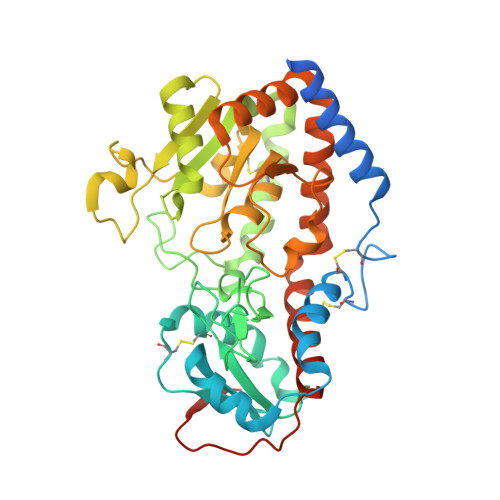
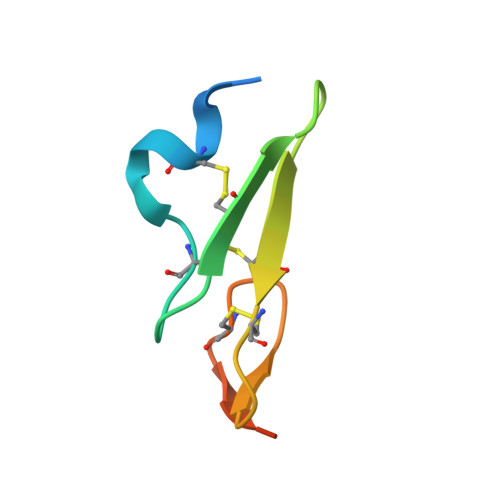
Structural analysis of Notch-regulating Rumi reveals basis for pathogenic mutations.

Yu, H.Takeuchi, H.Takeuchi, M.Liu, Q.Kantharia, J.Haltiwanger, R.S.Li, H.

(2016) Nat Chem Biol 12: 735-740

  • DOI: https://doi.org/10.1038/nchembio.2135
  • Primary Citation of Related Structures:  
    5F84, 5F85, 5F86, 5F87

  • PubMed Abstract: 

    Rumi O-glucosylates the EGF repeats of a growing list of proteins essential in metazoan development, including Notch. Rumi is essential for Notch signaling, and Rumi dysregulation is linked to several human diseases. Despite Rumi's critical roles, it is unknown how Rumi glucosylates a serine of many but not all EGF repeats. Here we report crystal structures of Drosophila Rumi as binary and ternary complexes with a folded EGF repeat and/or donor substrates. These structures provide insights into the catalytic mechanism and show that Rumi recognizes structural signatures of the EGF motif, the U-shaped consensus sequence, C-X-S-X-(P/A)-C and a conserved hydrophobic region. We found that five Rumi mutations identified in cancers and Dowling-Degos disease are clustered around the enzyme active site and adversely affect its activity. Our study suggests that loss of Rumi activity may underlie these diseases, and the mechanistic insights may facilitate the development of modulators of Notch signaling.


  • Organizational Affiliation

    Biology Department, Brookhaven National Laboratory, Upton, New York, USA.


Macromolecules
Find similar proteins by:  (by identity cutoff)  |  3D Structure
Entity ID: 1
MoleculeChains Sequence LengthOrganismDetailsImage
O-glucosyltransferase rumi402Drosophila melanogasterMutation(s): 0 
Gene Names: rumiCG31152
EC: 2.4.1
UniProt
Find proteins for Q8T045 (Drosophila melanogaster)
Explore Q8T045 
Go to UniProtKB:  Q8T045
Entity Groups  
Sequence Clusters30% Identity50% Identity70% Identity90% Identity95% Identity100% Identity
UniProt GroupQ8T045
Sequence Annotations
Expand
  • Reference Sequence
Find similar proteins by:  (by identity cutoff)  |  3D Structure
Entity ID: 2
MoleculeChains Sequence LengthOrganismDetailsImage
Coagulation factor IX50Homo sapiensMutation(s): 0 
Gene Names: F9
EC: 3.4.21.22
UniProt & NIH Common Fund Data Resources
Find proteins for P00740 (Homo sapiens)
Explore P00740 
Go to UniProtKB:  P00740
PHAROS:  P00740
GTEx:  ENSG00000101981 
Entity Groups  
Sequence Clusters30% Identity50% Identity70% Identity90% Identity95% Identity100% Identity
UniProt GroupP00740
Sequence Annotations
Expand
  • Reference Sequence
Experimental Data & Validation

Experimental Data

  • Method: X-RAY DIFFRACTION
  • Resolution: 2.50 Å
  • R-Value Free: 0.239 
  • R-Value Work: 0.192 
  • R-Value Observed: 0.194 
  • Space Group: H 3 2
Unit Cell:
Length ( Å )Angle ( ˚ )
a = 271.016α = 90
b = 271.016β = 90
c = 47.427γ = 120
Software Package:
Software NamePurpose
REFMACrefinement
HKL-2000data scaling
HKL-2000data reduction
MOLREPphasing

Structure Validation

View Full Validation Report


Ligand Structure Quality Assessment 


Entry History 

Deposition Data

Revision History  (Full details and data files)

  • Version 1.0: 2016-07-20
    Type: Initial release
  • Version 1.1: 2018-04-18
    Changes: Data collection, Database references, Derived calculations
  • Version 1.2: 2020-07-29
    Type: Remediation
    Reason: Carbohydrate remediation
    Changes: Data collection, Derived calculations, Structure summary