5EZ2

Sandercyanin Fluorescent Protein (SFP)


Experimental Data Snapshot

  • Method: X-RAY DIFFRACTION
  • Resolution: 1.85 Å
  • R-Value Free: 0.212 
  • R-Value Work: 0.190 
  • R-Value Observed: 0.191 

wwPDB Validation   3D Report Full Report


Ligand Structure Quality Assessment 


This is version 1.2 of the entry. See complete history


Literature

Blue protein with red fluorescence

Ghosh, S.Yu, C.L.Ferraro, D.J.Sudha, S.Pal, S.K.Schaefer, W.F.Gibson, D.T.Ramaswamy, S.

(2016) Proc Natl Acad Sci U S A 113: 11513-11518

  • DOI: https://doi.org/10.1073/pnas.1525622113
  • Primary Citation of Related Structures:  
    5EZ2, 5F1E

  • PubMed Abstract: 

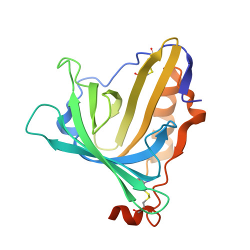
    The walleye (Sander vitreus) is a golden yellow fish that inhabits the Northern American lakes. The recent sightings of the blue walleye and the correlation of its sighting to possible increased UV radiation have been proposed earlier. The underlying molecular basis of its adaptation to increased UV radiation is the presence of a protein (Sandercyanin)-ligand complex in the mucus of walleyes. Degradation of heme by UV radiation results in the formation of Biliverdin IXα (BLA), the chromophore bound to Sandercyanin. We show that Sandercyanin is a monomeric protein that forms stable homotetramers on addition of BLA to the protein. A structure of the Sandercyanin-BLA complex, purified from the fish mucus, reveals a glycosylated protein with a lipocalin fold. This protein-ligand complex absorbs light in the UV region (λ max of 375 nm) and upon excitation at this wavelength emits in the red region (λ max of 675 nm). Unlike all other known biliverdin-bound fluorescent proteins, the chromophore is noncovalently bound to the protein. We provide here a molecular rationale for the observed spectral properties of Sandercyanin.


  • Organizational Affiliation

    National Center for Biological Sciences, Tata Institute of Fundamental Research, Bangalore, India 560065.


Macromolecules
Find similar proteins by:  (by identity cutoff)  |  3D Structure
Entity ID: 1
MoleculeChains Sequence LengthOrganismDetailsImage
Sandercyanin Fluorescent Protein
A, B
183Sander vitreusMutation(s): 0 
UniProt
Find proteins for A0A1D5B367 (Sander vitreus)
Explore A0A1D5B367 
Go to UniProtKB:  A0A1D5B367
Entity Groups  
Sequence Clusters30% Identity50% Identity70% Identity90% Identity95% Identity100% Identity
UniProt GroupA0A1D5B367
Sequence Annotations
Expand
  • Reference Sequence
Experimental Data & Validation

Experimental Data

  • Method: X-RAY DIFFRACTION
  • Resolution: 1.85 Å
  • R-Value Free: 0.212 
  • R-Value Work: 0.190 
  • R-Value Observed: 0.191 
  • Space Group: P 63 2 2
Unit Cell:
Length ( Å )Angle ( ˚ )
a = 159.266α = 90
b = 159.266β = 90
c = 84.157γ = 120
Software Package:
Software NamePurpose
PHENIXrefinement
HKL-2000data processing
HKL-2000data scaling
PHASERphasing
iMOSFLMdata reduction

Structure Validation

View Full Validation Report


Ligand Structure Quality Assessment 


Entry History 

Deposition Data

Revision History  (Full details and data files)

  • Version 1.0: 2016-09-28
    Type: Initial release
  • Version 1.1: 2016-10-26
    Changes: Database references
  • Version 1.2: 2023-11-08
    Changes: Data collection, Database references, Derived calculations, Refinement description