5EUJ

PYRUVATE DECARBOXYLASE FROM ZYMOBACTER PALMAE


Experimental Data Snapshot

  • Method: X-RAY DIFFRACTION
  • Resolution: 2.15 Å
  • R-Value Free: 0.220 
  • R-Value Work: 0.186 
  • R-Value Observed: 0.188 

wwPDB Validation   3D Report Full Report


Ligand Structure Quality Assessment 


This is version 1.3 of the entry. See complete history


Literature

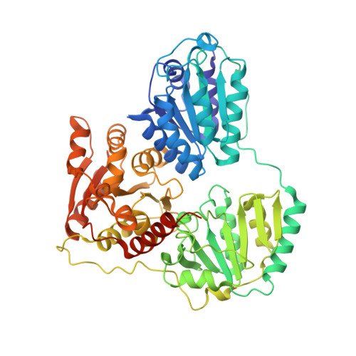
Crystal structure of pyruvate decarboxylase from Zymobacter palmae.

Buddrus, L.Andrews, E.S.Leak, D.J.Danson, M.J.Arcus, V.L.Crennell, S.J.

(2016) Acta Crystallogr F Struct Biol Commun 72: 700-706

  • DOI: https://doi.org/10.1107/S2053230X16012012
  • Primary Citation of Related Structures:  
    5EUJ

  • PubMed Abstract: 

    Pyruvate decarboxylase (PDC; EC 4.1.1.1) is a thiamine pyrophosphate- and Mg(2+) ion-dependent enzyme that catalyses the non-oxidative decarboxylation of pyruvate to acetaldehyde and carbon dioxide. It is rare in bacteria, but is a key enzyme in homofermentative metabolism, where ethanol is the major product. Here, the previously unreported crystal structure of the bacterial pyruvate decarboxylase from Zymobacter palmae is presented. The crystals were shown to diffract to 2.15 Å resolution. They belonged to space group P21, with unit-cell parameters a = 204.56, b = 177.39, c = 244.55 Å and Rr.i.m. = 0.175 (0.714 in the highest resolution bin). The structure was solved by molecular replacement using PDB entry 2vbi as a model and the final R values were Rwork = 0.186 (0.271 in the highest resolution bin) and Rfree = 0.220 (0.300 in the highest resolution bin). Each of the six tetramers is a dimer of dimers, with each monomer sharing its thiamine pyrophosphate across the dimer interface, and some contain ethylene glycol mimicking the substrate pyruvate in the active site. Comparison with other bacterial PDCs shows a correlation of higher thermostability with greater tetramer interface area and number of interactions.


  • Organizational Affiliation

    Department of Biology and Biochemistry, University of Bath, Claverton Down, Bath BA2 7AY, England.


Macromolecules
Find similar proteins by:  (by identity cutoff)  |  3D Structure
Entity ID: 1
MoleculeChains Sequence LengthOrganismDetailsImage
Pyruvate decarboxylase
A, B, C, D, E
A, B, C, D, E, F, G, H, I, J, K, L, M, N, O, P, Q, R, S, T, U, V, W, X
573Zymobacter palmaeMutation(s): 0 
Gene Names: pdc
EC: 4.1.1.1
UniProt
Find proteins for Q8KTX6 (Zymobacter palmae)
Explore Q8KTX6 
Go to UniProtKB:  Q8KTX6
Entity Groups  
Sequence Clusters30% Identity50% Identity70% Identity90% Identity95% Identity100% Identity
UniProt GroupQ8KTX6
Sequence Annotations
Expand
  • Reference Sequence
Small Molecules
Ligands 3 Unique
IDChains Name / Formula / InChI Key2D Diagram3D Interactions
TPP
Query on TPP

Download Ideal Coordinates CCD File 
AB [auth M]
CA [auth B]
CB [auth N]
EA [auth C]
FB [auth O]
AB [auth M],
CA [auth B],
CB [auth N],
EA [auth C],
FB [auth O],
GA [auth D],
HB [auth P],
IA [auth E],
JB [auth Q],
LA [auth F],
MB [auth R],
NA [auth G],
PA [auth H],
PB [auth S],
RA [auth I],
RB [auth T],
TB [auth U],
UA [auth J],
VB [auth V],
WA [auth K],
XB [auth W],
YA [auth L],
Z [auth A],
ZB [auth X]
THIAMINE DIPHOSPHATE
C12 H19 N4 O7 P2 S
AYEKOFBPNLCAJY-UHFFFAOYSA-O
EDO
Query on EDO

Download Ideal Coordinates CCD File 
AA [auth A]
DB [auth N]
JA [auth F]
KB [auth Q]
NB [auth R]
AA [auth A],
DB [auth N],
JA [auth F],
KB [auth Q],
NB [auth R],
SA [auth I]
1,2-ETHANEDIOL
C2 H6 O2
LYCAIKOWRPUZTN-UHFFFAOYSA-N
MG
Query on MG

Download Ideal Coordinates CCD File 
BA [auth B]
BB [auth N]
DA [auth C]
EB [auth O]
FA [auth D]
BA [auth B],
BB [auth N],
DA [auth C],
EB [auth O],
FA [auth D],
GB [auth P],
HA [auth E],
IB [auth Q],
KA [auth F],
LB [auth R],
MA [auth G],
OA [auth H],
OB [auth S],
QA [auth I],
QB [auth T],
SB [auth U],
TA [auth J],
UB [auth V],
VA [auth K],
WB [auth W],
XA [auth L],
Y [auth A],
YB [auth X],
ZA [auth M]
MAGNESIUM ION
Mg
JLVVSXFLKOJNIY-UHFFFAOYSA-N
Experimental Data & Validation

Experimental Data

  • Method: X-RAY DIFFRACTION
  • Resolution: 2.15 Å
  • R-Value Free: 0.220 
  • R-Value Work: 0.186 
  • R-Value Observed: 0.188 
  • Space Group: P 1 21 1
Unit Cell:
Length ( Å )Angle ( ˚ )
a = 204.557α = 90
b = 177.39β = 112.94
c = 244.553γ = 90
Software Package:
Software NamePurpose
REFMACrefinement
PDB_EXTRACTdata extraction
Aimlessdata scaling
iMOSFLMdata reduction
BALBESphasing

Structure Validation

View Full Validation Report


Ligand Structure Quality Assessment 


Entry History 

Deposition Data

Revision History  (Full details and data files)

  • Version 1.0: 2016-09-14
    Type: Initial release
  • Version 1.1: 2016-09-28
    Changes: Database references
  • Version 1.2: 2017-07-12
    Changes: Structure summary
  • Version 1.3: 2024-01-10
    Changes: Data collection, Database references, Derived calculations, Refinement description