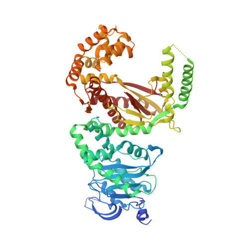
5DKT

N-terminal His tagged apPOL exonuclease mutant


Experimental Data Snapshot

  • Method: X-RAY DIFFRACTION
  • Resolution: 2.90 Å
  • R-Value Free: 0.299 
  • R-Value Work: 0.265 
  • R-Value Observed: 0.268 

wwPDB Validation   3D Report Full Report


This is version 1.3 of the entry. See complete history


Literature

Crystal Structure of the Apicoplast DNA Polymerase from Plasmodium falciparum: The First Look at a Plastidic A-Family DNA Polymerase.

Milton, M.E.Choe, J.Y.Honzatko, R.B.Nelson, S.W.

(2016) J Mol Biol 428: 3920-3934

  • DOI: https://doi.org/10.1016/j.jmb.2016.07.016
  • Primary Citation of Related Structures:  
    5DKT, 5DKU

  • PubMed Abstract: 

    Plasmodium falciparum, the primary cause of malaria, contains a non-photosynthetic plastid called the apicoplast. The apicoplast exists in most members of the phylum Apicomplexa and has its own genome along with organelle-specific enzymes for its replication. The only DNA polymerase found in the apicoplast (apPOL) was putatively acquired through horizontal gene transfer from a bacteriophage and is classified as an atypical A-family polymerase. Here, we present its crystal structure at a resolution of 2.9Å. P. falciparum apPOL, the first structural representative of a plastidic A-family polymerase, diverges from typical A-family members in two of three previously identified signature motifs and in a region not implicated by sequence. Moreover, apPOL has an additional N-terminal subdomain, the absence of which severely diminishes its 3' to 5' exonuclease activity. A compound known to be toxic to Plasmodium is a potent inhibitor of apPOL, suggesting that apPOL is a viable drug target. The structure provides new insights into the structural diversity of A-family polymerases and may facilitate structurally guided antimalarial drug design.


  • Organizational Affiliation

    Department of Biochemistry, Biophysics, and Molecular Biology, Iowa State University, Ames, IA 50011, USA.


Macromolecules
Find similar proteins by:  (by identity cutoff)  |  3D Structure
Entity ID: 1
MoleculeChains Sequence LengthOrganismDetailsImage
Prex DNA polymerase648Plasmodium falciparum 3D7Mutation(s): 2 
Gene Names: POM1PF14_0112
EC: 2.7.7.7
UniProt
Find proteins for Q8ILY1 (Plasmodium falciparum (isolate 3D7))
Explore Q8ILY1 
Go to UniProtKB:  Q8ILY1
Entity Groups  
Sequence Clusters30% Identity50% Identity70% Identity90% Identity95% Identity100% Identity
UniProt GroupQ8ILY1
Sequence Annotations
Expand
  • Reference Sequence
Small Molecules
Ligands 2 Unique
IDChains Name / Formula / InChI Key2D Diagram3D Interactions
SO4
Query on SO4

Download Ideal Coordinates CCD File 
B [auth A]
C [auth A]
D [auth A]
E [auth A]
F [auth A]
B [auth A],
C [auth A],
D [auth A],
E [auth A],
F [auth A],
G [auth A],
H [auth A],
I [auth A],
J [auth A]
SULFATE ION
O4 S
QAOWNCQODCNURD-UHFFFAOYSA-L
NA
Query on NA

Download Ideal Coordinates CCD File 
K [auth A]SODIUM ION
Na
FKNQFGJONOIPTF-UHFFFAOYSA-N
Experimental Data & Validation

Experimental Data

  • Method: X-RAY DIFFRACTION
  • Resolution: 2.90 Å
  • R-Value Free: 0.299 
  • R-Value Work: 0.265 
  • R-Value Observed: 0.268 
  • Space Group: P 65 2 2
Unit Cell:
Length ( Å )Angle ( ˚ )
a = 141.775α = 90
b = 141.775β = 90
c = 148.759γ = 120
Software Package:
Software NamePurpose
PHENIXrefinement
HKL-3000data reduction
HKL-3000data scaling
MR-Rosettaphasing

Structure Validation

View Full Validation Report


Entry History & Funding Information

Deposition Data


Funding OrganizationLocationGrant Number
Iowa State University College of Liberal Arts and SciencesUnited States--

Revision History  (Full details and data files)

  • Version 1.0: 2016-08-10
    Type: Initial release
  • Version 1.1: 2016-08-17
    Changes: Database references
  • Version 1.2: 2016-10-19
    Changes: Database references
  • Version 1.3: 2023-09-27
    Changes: Data collection, Database references, Derived calculations, Refinement description