5DK8

Human ubiquitin in the P1 space group


Experimental Data Snapshot

  • Method: X-RAY DIFFRACTION
  • Resolution: 1.32 Å
  • R-Value Free: 0.201 
  • R-Value Work: 0.178 
  • R-Value Observed: 0.180 

wwPDB Validation   3D Report Full Report


This is version 1.3 of the entry. See complete history


Literature

New crystal form of human ubiquitin in the presence of magnesium.

Camara-Artigas, A.Plaza-Garrido, M.Martinez-Rodriguez, S.Bacarizo, J.

(2016) Acta Crystallogr F Struct Biol Commun 72: 29-35

  • DOI: https://doi.org/10.1107/S2053230X15023390
  • Primary Citation of Related Structures:  
    5DK8

  • PubMed Abstract: 

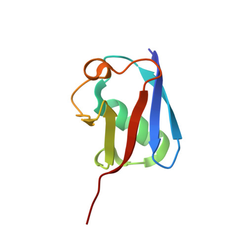
    Ubiquitin is a small globular protein that has a considerable number of lysine residues on its surface. This results in a high surface entropy that precludes the formation of crystal-packing interactions. To date, only a few structures of the native form of ubiquitin have been solved, and most of the crystals that led to these structures were obtained in the presence of different divalent metal cations. In this work, a new crystallographic structure of human ubiquitin solved from crystals grown in the presence of magnesium is presented. The crystals belonged to a triclinic space group, with unit-cell parameters a = 29.96, b = 30.18, c = 41.41 Å, α = 88.52, β = 79.12, γ = 67.37°. The crystal lattice is composed of stacked layers of human ubiquitin molecules with a large hydrophobic interface and a smaller polar interface in which the magnesium ion lies at the junction between adjacent layers in the crystal. The metal ion appears in a hexa-aquo coordination, which is key to facilitating the crystallization of the protein.


  • Organizational Affiliation

    Department of Chemistry and Physics, University of Almeria, Agrifood Campus of International Excellence (ceiA3), Carretera de Sacramento (s/n), 04120 Almeria, Spain.


Macromolecules
Find similar proteins by:  (by identity cutoff)  |  3D Structure
Entity ID: 1
MoleculeChains Sequence LengthOrganismDetailsImage
Polyubiquitin-B
A, B
76Homo sapiensMutation(s): 0 
Gene Names: UBB
UniProt & NIH Common Fund Data Resources
Find proteins for P0CG47 (Homo sapiens)
Explore P0CG47 
Go to UniProtKB:  P0CG47
PHAROS:  P0CG47
GTEx:  ENSG00000170315 
Entity Groups  
Sequence Clusters30% Identity50% Identity70% Identity90% Identity95% Identity100% Identity
UniProt GroupP0CG47
Sequence Annotations
Expand
  • Reference Sequence
Small Molecules
Ligands 1 Unique
IDChains Name / Formula / InChI Key2D Diagram3D Interactions
MG
Query on MG

Download Ideal Coordinates CCD File 
C [auth A]MAGNESIUM ION
Mg
JLVVSXFLKOJNIY-UHFFFAOYSA-N
Experimental Data & Validation

Experimental Data

  • Method: X-RAY DIFFRACTION
  • Resolution: 1.32 Å
  • R-Value Free: 0.201 
  • R-Value Work: 0.178 
  • R-Value Observed: 0.180 
  • Space Group: P 1
Unit Cell:
Length ( Å )Angle ( ˚ )
a = 29.961α = 88.52
b = 30.182β = 79.12
c = 41.414γ = 67.37
Software Package:
Software NamePurpose
Aimlessdata scaling
PHASERphasing
PHENIXrefinement
PDB_EXTRACTdata extraction
autoPROCdata reduction

Structure Validation

View Full Validation Report


Entry History & Funding Information

Deposition Data


Funding OrganizationLocationGrant Number
Ministry of Economy and CompetitivenessSpainBIO2012-39922-C02-01/02

Revision History  (Full details and data files)

  • Version 1.0: 2015-12-16
    Type: Initial release
  • Version 1.1: 2016-01-13
    Changes: Database references
  • Version 1.2: 2016-01-20
    Changes: Database references
  • Version 1.3: 2024-01-10
    Changes: Data collection, Database references, Derived calculations, Refinement description