Structural insight for substrate tolerance to 2-deoxyribose-5-phosphate aldolase from the pathogen Streptococcus suis
Cao, T.-P., Kim, J.-S., Woo, M.-H., Choi, J.M., Jun, Y., Lee, K.H., Lee, S.H.(2016) J Microbiol 54: 311-321
- PubMed: 27033207
- DOI: https://doi.org/10.1007/s12275-016-6029-4
- Primary Citation of Related Structures:
5DBT, 5DBU - PubMed Abstract:
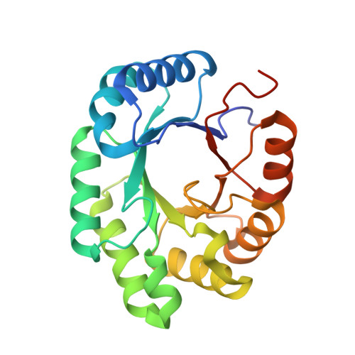
2-deoxyribose-5-phosphate aldolase (DERA) is a class I aldolase that catalyzes aldol condensation of two aldehydes in the active site, which is particularly germane in drug manufacture. Structural and biochemical studies have shown that the active site of DERA is typically loosely packed and displays broader substrate specificity despite sharing conserved folding architecture with other aldolases. The most distinctive structural feature of DERA compared to other aldolases is short and flexible C-terminal region. This region is also responsible for substrate recognition. Therefore, substrate tolerance may be related to the C-terminal structural features of DERA. Here, we determined the crystal structures of full length and C-terminal truncated DERA from Streptococcus suis (SsDERA). In common, both contained the typical (α/β)8 TIM-barrel fold of class I aldolases. Surprisingly, C-terminal truncation resulting in missing the last α9 and β8 secondary elements, allowed DERA to maintain activity comparable to the fulllength enzyme. Specifically, Arg186 and Ser205 residues at the C-terminus appeared mutually supplemental or less indispensible for substrate phosphate moiety recognition. Our results suggest that DERA might adopt a shorter C-terminal region than conventional aldolases during evolution pathway, resulting in a broader range of substrate tolerance through active site flexibility.
Organizational Affiliation:
Department of Cellular and Molecular Medicine, Chosun University School of Medicine, Gwangju, 501-759, Republic of Korea.