5D83

Crystal Structure of Ketosteroid Isomerase from Pseudomonas putida (pKSI); D40N, Y32(Cl-Y)


Experimental Data Snapshot

  • Method: X-RAY DIFFRACTION
  • Resolution: 1.70 Å
  • R-Value Free: 0.248 
  • R-Value Work: 0.196 
  • R-Value Observed: 0.199 

wwPDB Validation   3D Report Full Report


This is version 1.3 of the entry. See complete history


Literature

Dissecting Proton Delocalization in an Enzyme's Hydrogen Bond Network with Unnatural Amino Acids.

Wu, Y.Fried, S.D.Boxer, S.G.

(2015) Biochemistry 54: 7110-7119

  • DOI: https://doi.org/10.1021/acs.biochem.5b00958
  • Primary Citation of Related Structures:  
    5D81, 5D82, 5D83

  • PubMed Abstract: 

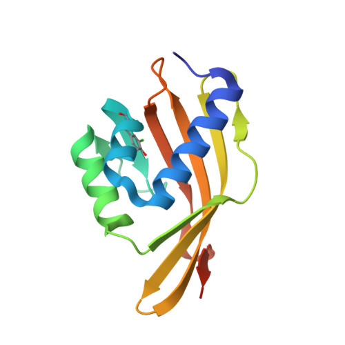
    Extended hydrogen bond networks are a common structural motif of enzymes. A recent analysis proposed quantum delocalization of protons as a feature present in the hydrogen bond network spanning a triad of tyrosines (Y(16), Y(32), and Y(57)) in the active site of ketosteroid isomerase (KSI), contributing to its unusual acidity and large isotope shift. In this study, we utilized amber suppression to substitute each tyrosine residue with 3-chlorotyrosine to test the delocalization model and the proton affinity balance in the triad. X-ray crystal structures of each variant demonstrated that the structure, notably the O-O distances within the triad, was unaffected by 3-chlorotyrosine substitutions. The changes in the cluster's acidity and the acidity's isotope dependence in these variants were assessed via UV-vis spectroscopy and the proton sharing pattern among individual residues with (13)C nuclear magnetic resonance. Our data show pKa detuning at each triad residue alters the proton delocalization behavior in the H-bond network. The extra stabilization energy necessary for the unusual acidity mainly comes from the strong interactions between Y(57) and Y(16). This is further enabled by Y(32), which maintains the right geometry and matched proton affinity in the triad. This study provides a rich picture of the energetics of the hydrogen bond network in enzymes for further model refinement.


  • Organizational Affiliation

    Department of Chemistry, Stanford University , Stanford, California 94305-5012, United States.


Macromolecules
Find similar proteins by:  (by identity cutoff)  |  3D Structure
Entity ID: 1
MoleculeChains Sequence LengthOrganismDetailsImage
Delta(5)-3-ketosteroid isomerase
A, B
135Pseudomonas putidaMutation(s): 1 
Gene Names: ksi
EC: 5.3.3.1
UniProt
Find proteins for P07445 (Pseudomonas putida)
Explore P07445 
Go to UniProtKB:  P07445
Entity Groups  
Sequence Clusters30% Identity50% Identity70% Identity90% Identity95% Identity100% Identity
UniProt GroupP07445
Sequence Annotations
Expand
  • Reference Sequence
Small Molecules
Modified Residues  1 Unique
IDChains TypeFormula2D DiagramParent
3CT
Query on 3CT
A, B
L-PEPTIDE LINKINGC9 H10 Cl N O3TYR
Experimental Data & Validation

Experimental Data

  • Method: X-RAY DIFFRACTION
  • Resolution: 1.70 Å
  • R-Value Free: 0.248 
  • R-Value Work: 0.196 
  • R-Value Observed: 0.199 
  • Space Group: P 21 21 21
Unit Cell:
Length ( Å )Angle ( ˚ )
a = 34.9α = 90
b = 72.76β = 90
c = 94.95γ = 90
Software Package:
Software NamePurpose
PHENIXrefinement
SCALAdata scaling
PDB_EXTRACTdata extraction
Cootmodel building
PHASERphasing

Structure Validation

View Full Validation Report


Entry History & Funding Information

Deposition Data


Funding OrganizationLocationGrant Number
National Institutes of Health/National Institute of General Medical Sciences (NIH/NIGMS)United States--

Revision History  (Full details and data files)

  • Version 1.0: 2015-12-02
    Type: Initial release
  • Version 1.1: 2015-12-23
    Changes: Database references
  • Version 1.2: 2018-05-23
    Changes: Author supporting evidence, Data collection, Database references, Derived calculations
  • Version 1.3: 2019-12-25
    Changes: Author supporting evidence