5D0U

Crystal structure of the RNA-helicase Prp43 from Chaetomium thermophilum bound to ADP


Experimental Data Snapshot

  • Method: X-RAY DIFFRACTION
  • Resolution: 2.92 Å
  • R-Value Free: 0.231 
  • R-Value Work: 0.190 
  • R-Value Observed: 0.193 

wwPDB Validation   3D Report Full Report


Ligand Structure Quality Assessment 


This is version 1.3 of the entry. See complete history


Literature

Structural and functional analysis of the RNA helicase Prp43 from the thermophilic eukaryote Chaetomium thermophilum.

Tauchert, M.J.Fourmann, J.B.Christian, H.Luhrmann, R.Ficner, R.

(2016) Acta Crystallogr F Struct Biol Commun 72: 112-120

  • DOI: https://doi.org/10.1107/S2053230X15024498
  • Primary Citation of Related Structures:  
    5D0U

  • PubMed Abstract: 

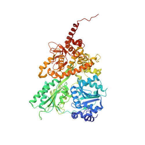
    RNA helicases are indispensable for all organisms in each domain of life and have implications in numerous cellular processes. The DEAH-box RNA helicase Prp43 is involved in pre-mRNA splicing as well as rRNA maturation. Here, the crystal structure of Chaetomium thermophilum Prp43 at 2.9 Å resolution is revealed. Furthermore, it is demonstrated that Prp43 from C. thermophilum is capable of functionally replacing its orthologue from Saccharomyces cerevisiae in spliceosomal disassembly assays.


  • Organizational Affiliation

    Department of Molecular Structural Biology, Institute for Microbiology and Genetics, GZMB, Georg-August-Universität Göttingen, Justus-von-Liebig Weg 11, 37077 Göttingen, Germany.


Macromolecules
Find similar proteins by:  (by identity cutoff)  |  3D Structure
Entity ID: 1
MoleculeChains Sequence LengthOrganismDetailsImage
Pre-mRNA-splicing factor ATP-dependent RNA helicase PRP43714Thermochaetoides thermophila DSM 1495Mutation(s): 0 
Gene Names: CTHT_0005780
UniProt
Find proteins for G0RY84 (Chaetomium thermophilum (strain DSM 1495 / CBS 144.50 / IMI 039719))
Explore G0RY84 
Go to UniProtKB:  G0RY84
Entity Groups  
Sequence Clusters30% Identity50% Identity70% Identity90% Identity95% Identity100% Identity
UniProt GroupG0RY84
Sequence Annotations
Expand
  • Reference Sequence
Small Molecules
Ligands 3 Unique
IDChains Name / Formula / InChI Key2D Diagram3D Interactions
ADP
Query on ADP

Download Ideal Coordinates CCD File 
B [auth A]ADENOSINE-5'-DIPHOSPHATE
C10 H15 N5 O10 P2
XTWYTFMLZFPYCI-KQYNXXCUSA-N
DMS
Query on DMS

Download Ideal Coordinates CCD File 
D [auth A]
E [auth A]
F [auth A]
G [auth A]
H [auth A]
D [auth A],
E [auth A],
F [auth A],
G [auth A],
H [auth A],
I [auth A],
J [auth A],
K [auth A],
L [auth A]
DIMETHYL SULFOXIDE
C2 H6 O S
IAZDPXIOMUYVGZ-UHFFFAOYSA-N
MG
Query on MG

Download Ideal Coordinates CCD File 
C [auth A]MAGNESIUM ION
Mg
JLVVSXFLKOJNIY-UHFFFAOYSA-N
Experimental Data & Validation

Experimental Data

  • Method: X-RAY DIFFRACTION
  • Resolution: 2.92 Å
  • R-Value Free: 0.231 
  • R-Value Work: 0.190 
  • R-Value Observed: 0.193 
  • Space Group: P 65
Unit Cell:
Length ( Å )Angle ( ˚ )
a = 152.211α = 90
b = 152.211β = 90
c = 92.766γ = 120
Software Package:
Software NamePurpose
PHENIXrefinement
XDSdata reduction
XSCALEdata scaling
PHASERphasing

Structure Validation

View Full Validation Report


Ligand Structure Quality Assessment 


Entry History & Funding Information

Deposition Data


Funding OrganizationLocationGrant Number
German Research FoundationGermanySFB 860

Revision History  (Full details and data files)

  • Version 1.0: 2016-02-10
    Type: Initial release
  • Version 1.1: 2016-07-27
    Changes: Database references
  • Version 1.2: 2017-09-06
    Changes: Author supporting evidence
  • Version 1.3: 2024-01-10
    Changes: Data collection, Database references, Refinement description