5CY0

Total Chemical Synthesis, Covalent Structure Verification, and X-ray Structure of Bioactive Ts3 Toxin by Racemic Protein Crystallography


Experimental Data Snapshot

  • Method: X-RAY DIFFRACTION
  • Resolution: 1.93 Å
  • R-Value Free: 0.304 
  • R-Value Work: 0.256 
  • R-Value Observed: 0.259 

wwPDB Validation   3D Report Full Report


This is version 1.6 of the entry. See complete history


Literature

Elucidation of the Covalent and Tertiary Structures of Biologically Active Ts3 Toxin.

Dang, B.Kubota, T.Mandal, K.Correa, A.M.Bezanilla, F.Kent, S.B.

(2016) Angew Chem Int Ed Engl 55: 8639-8642

  • DOI: https://doi.org/10.1002/anie.201603420
  • Primary Citation of Related Structures:  
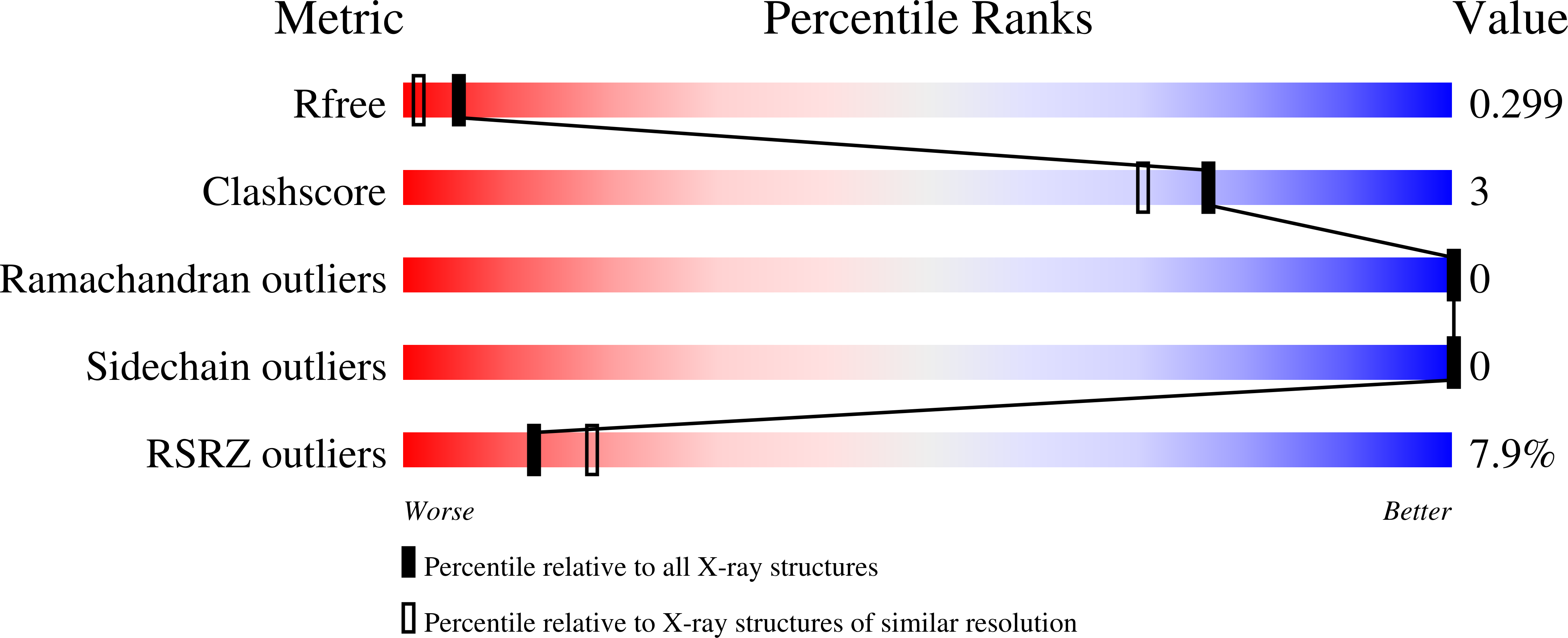
    5CY0

  • PubMed Abstract: 

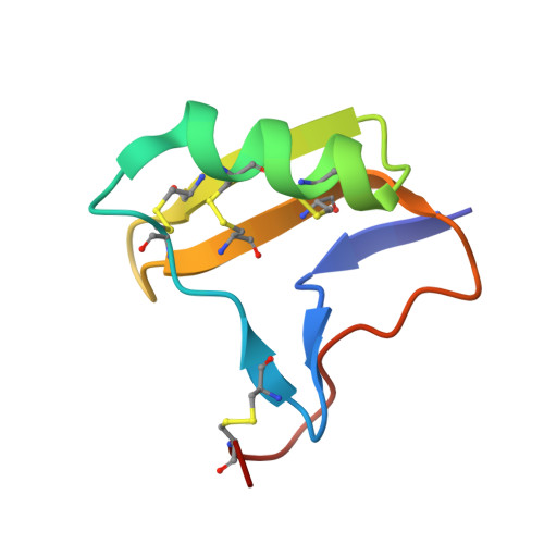
    Ts3 is an alpha scorpion toxin from the venom of the Brazilian scorpion Tityus serrulatus. Ts3 binds to the domain IV voltage sensor of voltage-gated sodium channels (Nav ) and slows down their fast inactivation. The covalent structure of the Ts3 toxin is uncertain, and the structure of the folded protein molecule is unknown. Herein, we report the total chemical synthesis of four candidate Ts3 toxin protein molecules and the results of structure-activity studies that enabled us to establish the covalent structure of biologically active Ts3 toxin. We also report the synthesis of the mirror image form of the Ts3 protein molecule, and the use of racemic protein crystallography to determine the folded (tertiary) structure of biologically active Ts3 toxin by X-ray diffraction.


  • Organizational Affiliation

    Department of Chemistry, University of Chicago, Chicago, IL, 60637, USA.


Macromolecules
Find similar proteins by:  (by identity cutoff)  |  3D Structure
Entity ID: 1
MoleculeChains Sequence LengthOrganismDetailsImage
Ts3 Toxin64Tityus serrulatusMutation(s): 0 
UniProt
Find proteins for P01496 (Tityus serrulatus)
Explore P01496 
Go to UniProtKB:  P01496
Entity Groups  
Sequence Clusters30% Identity50% Identity70% Identity90% Identity95% Identity100% Identity
UniProt GroupP01496
Sequence Annotations
Expand
  • Reference Sequence
Experimental Data & Validation

Experimental Data

  • Method: X-RAY DIFFRACTION
  • Resolution: 1.93 Å
  • R-Value Free: 0.304 
  • R-Value Work: 0.256 
  • R-Value Observed: 0.259 
  • Space Group: P -1
Unit Cell:
Length ( Å )Angle ( ˚ )
a = 19.322α = 101.17
b = 30.406β = 91.26
c = 52.468γ = 98.61
Software Package:
Software NamePurpose
PHENIXrefinement
PHASERphasing
XDSdata reduction
XDSdata scaling

Structure Validation

View Full Validation Report


Entry History & Funding Information

Deposition Data


Funding OrganizationLocationGrant Number
National Institutes of Health/National Institute of General Medical Sciences (NIH/NIGMS)United StatesU54 GM087519
National Institutes of Health/National Institute of General Medical Sciences (NIH/NIGMS)United StatesR01- GM030376
American Heart AssociationUnited States13POST14800031
National Institutes of Health/National Institute of General Medical Sciences (NIH/NIGMS)United StatesP41 GM103403

Revision History  (Full details and data files)

  • Version 1.0: 2016-06-01
    Type: Initial release
  • Version 1.1: 2016-06-15
    Changes: Database references
  • Version 1.2: 2016-07-27
    Changes: Database references
  • Version 1.3: 2017-09-13
    Changes: Author supporting evidence, Database references, Derived calculations
  • Version 1.4: 2018-01-17
    Changes: Author supporting evidence
  • Version 1.5: 2019-12-25
    Changes: Author supporting evidence
  • Version 1.6: 2023-09-27
    Changes: Data collection, Database references, Refinement description