5CVM

USP46~ubiquitin BEA covalent complex


Experimental Data Snapshot

  • Method: X-RAY DIFFRACTION
  • Resolution: 1.90 Å
  • R-Value Free: 0.197 
  • R-Value Work: 0.176 
  • R-Value Observed: 0.177 

wwPDB Validation   3D Report Full Report


This is version 1.1 of the entry. See complete history


Literature

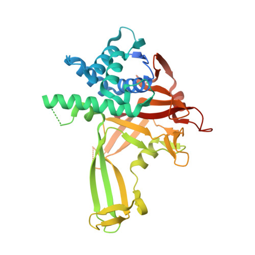
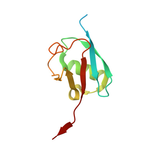
Structural Insights into WD-Repeat 48 Activation of Ubiquitin-Specific Protease 46.

Yin, J.Schoeffler, A.J.Wickliffe, K.Newton, K.Starovasnik, M.A.Dueber, E.C.Harris, S.F.

(2015) Structure 23: 2043-2054

  • DOI: https://doi.org/10.1016/j.str.2015.08.010
  • Primary Citation of Related Structures:  
    5CVL, 5CVM, 5CVN, 5CVO

  • PubMed Abstract: 

    Protein ubiquitination patterns are an important component of cellular signaling. The WD-repeat protein WDR48 (USP1-associated factor UAF-1) stimulates activity of ubiquitin-specific proteases USP1, USP12, and USP46. To understand how WDR48 exerts its effect on the USP scaffold, we determined structures of the ternary WDR48:USP46:ubiquitin complex. WDR48 interacts with the USP46 fingers subdomain via a relatively small, highly polar surface on the top center of the WDR48 β propeller. In addition, WDR48 has a novel ancillary domain and a C-terminal SUMO-like domain encircling the USP46-bound ubiquitin. Mutation of residues involved in the WDR48:USP46 interaction abrogated both binding and deubiquitinase activity of the complex. An analogous mutation in USP1 similarly blocked WDR48-dependent activation. Our data suggest a possible mechanism of deubiquitinase stimulation via stabilization and prolonged residence time of substrate. The unprecedented mode of interaction between the USP fingers domain and the WD-repeat β propeller serves as a prototypical example for this family of deubiquitinases.


  • Organizational Affiliation

    Department of Structural Biology, Genentech, Inc., 1 DNA Way, South San Francisco, CA 94080, USA.


Macromolecules
Find similar proteins by:  (by identity cutoff)  |  3D Structure
Entity ID: 1
MoleculeChains Sequence LengthOrganismDetailsImage
Ubiquitin carboxyl-terminal hydrolase 46358Homo sapiensMutation(s): 0 
Gene Names: USP46
EC: 3.4.19.12
UniProt & NIH Common Fund Data Resources
Find proteins for P62068 (Homo sapiens)
Explore P62068 
Go to UniProtKB:  P62068
PHAROS:  P62068
GTEx:  ENSG00000109189 
Entity Groups  
Sequence Clusters30% Identity50% Identity70% Identity90% Identity95% Identity100% Identity
UniProt GroupP62068
Sequence Annotations
Expand
  • Reference Sequence
Find similar proteins by:  (by identity cutoff)  |  3D Structure
Entity ID: 2
MoleculeChains Sequence LengthOrganismDetailsImage
Polyubiquitin-B96Homo sapiensMutation(s): 0 
Gene Names: UBB
UniProt & NIH Common Fund Data Resources
Find proteins for P0CG47 (Homo sapiens)
Explore P0CG47 
Go to UniProtKB:  P0CG47
PHAROS:  P0CG47
GTEx:  ENSG00000170315 
Entity Groups  
Sequence Clusters30% Identity50% Identity70% Identity90% Identity95% Identity100% Identity
UniProt GroupP0CG47
Sequence Annotations
Expand
  • Reference Sequence
Small Molecules
Ligands 1 Unique
IDChains Name / Formula / InChI Key2D Diagram3D Interactions
ZN
Query on ZN

Download Ideal Coordinates CCD File 
C [auth A]ZINC ION
Zn
PTFCDOFLOPIGGS-UHFFFAOYSA-N
Experimental Data & Validation

Experimental Data

  • Method: X-RAY DIFFRACTION
  • Resolution: 1.90 Å
  • R-Value Free: 0.197 
  • R-Value Work: 0.176 
  • R-Value Observed: 0.177 
  • Space Group: C 2 2 21
Unit Cell:
Length ( Å )Angle ( ˚ )
a = 90.613α = 90
b = 105.12β = 90
c = 134.656γ = 90
Software Package:
Software NamePurpose
BUSTERrefinement
SCALEPACKdata scaling
PDB_EXTRACTdata extraction
DENZOdata reduction
PHASERphasing

Structure Validation

View Full Validation Report


Entry History 

Deposition Data

Revision History  (Full details and data files)

  • Version 1.0: 2015-10-07
    Type: Initial release
  • Version 1.1: 2015-11-11
    Changes: Database references