The Structure of a Type 3 Secretion System (T3SS) Ruler Protein Suggests a Molecular Mechanism for Needle Length Sensing.
Bergeron, J.R., Fernandez, L., Wasney, G.A., Vuckovic, M., Reffuveille, F., Hancock, R.E., Strynadka, N.C.(2016) J Biol Chem 291: 1676-1691
- PubMed: 26589798
- DOI: https://doi.org/10.1074/jbc.M115.684423
- Primary Citation of Related Structures:
5CUK, 5CUL - PubMed Abstract:
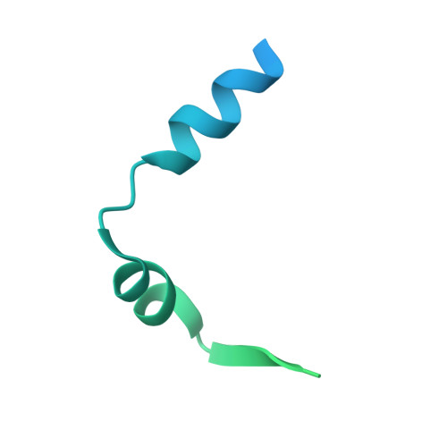
The type 3 secretion system (T3SS) and the bacterial flagellum are related pathogenicity-associated appendages found at the surface of many disease-causing bacteria. These appendages consist of long tubular structures that protrude away from the bacterial surface to interact with the host cell and/or promote motility. A proposed "ruler" protein tightly regulates the length of both the T3SS and the flagellum, but the molecular basis for this length control has remained poorly characterized and controversial. Using the Pseudomonas aeruginosa T3SS as a model system, we report the first structure of a T3SS ruler protein, revealing a "ball-and-chain" architecture, with a globular C-terminal domain (the ball) preceded by a long intrinsically disordered N-terminal polypeptide chain. The dimensions and stability of the globular domain do not support its potential passage through the inner lumen of the T3SS needle. We further demonstrate that a conserved motif at the N terminus of the ruler protein interacts with the T3SS autoprotease in the cytosolic side. Collectively, these data suggest a potential mechanism for needle length sensing by ruler proteins, whereby upon T3SS needle assembly, the ruler protein's N-terminal end is anchored on the cytosolic side, with the globular domain located on the extracellular end of the growing needle. Sequence analysis of T3SS and flagellar ruler proteins shows that this mechanism is probably conserved across systems.
Organizational Affiliation:
From the Department of Biochemistry and Molecular Biology,; the Centre for Blood Research, and.