Potent Inhibition of 3-Deoxy-d-arabinoheptulosonate-7-phosphate (DAHP) Synthase by DAHP Oxime, a Phosphate Group Mimic.
Balachandran, N., Heimhalt, M., Liuni, P., To, F., Wilson, D.J., Junop, M.S., Berti, P.J.(2016) Biochemistry 55: 6617-6629
- PubMed: 27933795
- DOI: https://doi.org/10.1021/acs.biochem.6b00930
- Primary Citation of Related Structures:
5CKS - PubMed Abstract:
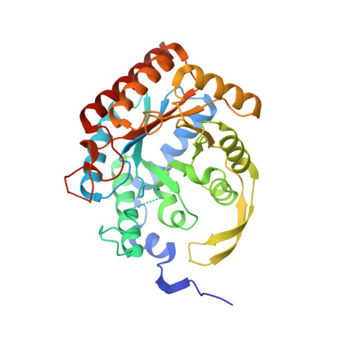
3-Deoxy-d-arabinoheptulosonate-7-phosphate (DAHP) synthase catalyzes the first step in the shikimate pathway. It catalyzes an aldol-like reaction of phosphoenolpyruvate (PEP) with erythrose 4-phosphate (E4P) to form DAHP. The kinetic mechanism was rapid equilibrium sequential ordered ter ter, with the essential divalent metal ion, Mn 2+ , binding first, followed by PEP and E4P. DAHP oxime, in which an oxime group replaces the keto oxygen, was a potent inhibitor, with K i = 1.5 ± 0.4 μM, though with residual activity at high inhibitor concentrations. It displayed slow-binding inhibition with a residence time, t R , of 83 min. The crystal structure revealed that the oxime functional group, combined with two crystallographic waters, bound at the same location in the catalytic center as the phosphate group of the tetrahedral intermediate. DAHP synthase has a dimer-of-dimers homotetrameric structure, and DAHP oxime bound to only one subunit of each tight dimer. Inhibitor binding was competitive with respect to all three substrates in the subunits to which it bound. DAHP oxime did not overlap with the metal binding site, so the cause of their mutually exclusive binding was not clear. Similarly, there was no obvious structural reason for inhibitor binding in only two subunits; however, changes in global hydrogen/deuterium exchange showed large scale changes in protein dynamics upon inhibitor binding. The k cat value for the residual activity at high inhibitor concentrations was 3-fold lower, and the apparent K M,E4P value decreased at least 10-fold. This positive cooperativity of binding between DAHP oxime in subunits B and C, and E4P in subunits A and D appears to be the dominant cause for incomplete inhibition at high inhibitor concentrations. In spite of its lack of obvious structural similarity to phosphate, the oxime and crystallographic waters acted as a small, neutral phosphate mimic.
Organizational Affiliation:
Department of Chemistry, York University , Toronto, Ontario M3J 1P3, Canada.