5CFA

Crystal structures of Bbp from Staphylococcus aureus with peptide ligand


Experimental Data Snapshot

  • Method: X-RAY DIFFRACTION
  • Resolution: 1.45 Å
  • R-Value Free: 0.212 
  • R-Value Work: 0.178 
  • R-Value Observed: 0.180 

wwPDB Validation   3D Report Full Report


This is version 1.2 of the entry. See complete history


Literature

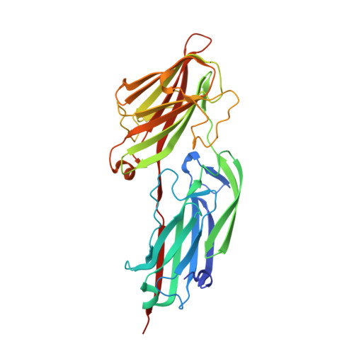
Crystal structures of Bbp from Staphylococcus aureus reveal the ligand binding mechanism with Fibrinogen alpha

Zhang, X.Y.Wu, M.Zhuo, W.Gu, J.K.Zhang, S.S.Ge, J.P.Yang, M.J.

(2015) Protein Cell 6: 757-766

  • DOI: https://doi.org/10.1007/s13238-015-0205-x
  • Primary Citation of Related Structures:  
    5CF3, 5CFA

  • PubMed Abstract: 

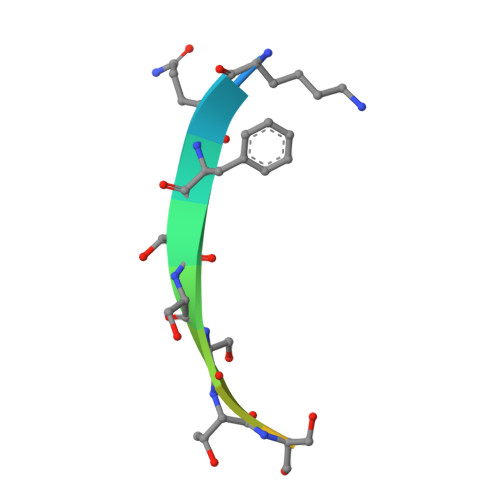
    Bone sialoprotein-binding protein (Bbp), a MSCRAMMs (Microbial Surface Components Recognizing Adhesive Matrix Molecules) family protein expressed on the surface of Staphylococcus aureus (S. aureus), mediates adherence to fibrinogen α (Fg α), a component in the extracellular matrix of the host cell and is important for infection and pathogenesis. In this study, we solved the crystal structures of apo-Bbp(273-598) and Bbp(273-598)-Fg α(561-575) complex at a resolution of 2.03 Å and 1.45 Å, respectively. Apo-Bbp(273-598) contained the ligand binding region N2 and N3 domains, both of which followed a DE variant IgG fold characterized by an additional D1 strand in N2 domain and D1' and D2' strands in N3 domain. The peptide mapped to the Fg α(561-575) bond to Bbp(273-598) on the open groove between the N2 and N3 domains. Strikingly, the disordered C-terminus in the apo-form reorganized into a highly-ordered loop and a β-strand G'' covering the ligand upon ligand binding. Bbp(Ala298-Gly301) in the N2 domain of the Bbp(273-598)-Fg α(561-575) complex, which is a loop in the apo-form, formed a short α-helix to interact tightly with the peptide. In addition, Bbp(Ser547-Gln561) in the N3 domain moved toward the binding groove to make contact directly with the peptide, while Bbp(Asp338-Gly355) and Bbp(Thr365-Tyr387) in N2 domain shifted their configurations to stabilize the reorganized C-terminus mainly through strong hydrogen bonds. Altogether, our results revealed the molecular basis for Bbp-ligand interaction and advanced our understanding of S. aureus infection process.


  • Organizational Affiliation

    Key Laboratory for Protein Sciences of Ministry of Education, Tsinghua-Peking Center for Life Sciences, School of Life Sciences, Tsinghua University, Beijing, 100084, China.


Macromolecules
Find similar proteins by:  (by identity cutoff)  |  3D Structure
Entity ID: 1
MoleculeChains Sequence LengthOrganismDetailsImage
Bone sialoprotein-binding protein
A, B
331Staphylococcus aureusMutation(s): 0 
Gene Names: bbp
UniProt
Find proteins for Q14U76 (Staphylococcus aureus)
Explore Q14U76 
Go to UniProtKB:  Q14U76
Entity Groups  
Sequence Clusters30% Identity50% Identity70% Identity90% Identity95% Identity100% Identity
UniProt GroupQ14U76
Sequence Annotations
Expand
  • Reference Sequence

Find similar proteins by:  Sequence   |   3D Structure  

Entity ID: 2
MoleculeChains Sequence LengthOrganismDetailsImage
Peptide from Fibrinogen alpha chainC [auth D],
D [auth C]
15Homo sapiensMutation(s): 0 
UniProt & NIH Common Fund Data Resources
Find proteins for P02671 (Homo sapiens)
Explore P02671 
Go to UniProtKB:  P02671
PHAROS:  P02671
GTEx:  ENSG00000171560 
Entity Groups  
Sequence Clusters30% Identity50% Identity70% Identity90% Identity95% Identity100% Identity
UniProt GroupP02671
Sequence Annotations
Expand
  • Reference Sequence
Small Molecules
Ligands 1 Unique
IDChains Name / Formula / InChI Key2D Diagram3D Interactions
MG
Query on MG

Download Ideal Coordinates CCD File 
E [auth A]MAGNESIUM ION
Mg
JLVVSXFLKOJNIY-UHFFFAOYSA-N
Experimental Data & Validation

Experimental Data

  • Method: X-RAY DIFFRACTION
  • Resolution: 1.45 Å
  • R-Value Free: 0.212 
  • R-Value Work: 0.178 
  • R-Value Observed: 0.180 
  • Space Group: P 1 21 1
Unit Cell:
Length ( Å )Angle ( ˚ )
a = 60.916α = 90
b = 74.961β = 102.91
c = 75.563γ = 90
Software Package:
Software NamePurpose
SCALEPACKdata scaling
PHENIXrefinement
PDB_EXTRACTdata extraction
HKL-2000data reduction
PHASERphasing

Structure Validation

View Full Validation Report


Entry History 

Deposition Data

Revision History  (Full details and data files)

  • Version 1.0: 2015-09-23
    Type: Initial release
  • Version 1.1: 2015-10-21
    Changes: Database references
  • Version 1.2: 2023-11-08
    Changes: Data collection, Database references, Derived calculations, Refinement description