5C9P

Crystal structure of recombinant PLL lectin complexed with L-fucose from Photorhabdus luminescens at 1.75 A resolution


Experimental Data Snapshot

  • Method: X-RAY DIFFRACTION
  • Resolution: 1.75 Å
  • R-Value Free: 0.181 
  • R-Value Work: 0.142 
  • R-Value Observed: 0.144 

wwPDB Validation   3D Report Full Report


Ligand Structure Quality Assessment 


This is version 1.4 of the entry. See complete history


Literature

A Novel Fucose-binding Lectin from Photorhabdus luminescens (PLL) with an Unusual Heptabladed beta-Propeller Tetrameric Structure.

Kumar, A.Sykorova, P.Demo, G.Dobes, P.Hyrsl, P.Wimmerova, M.

(2016) J Biol Chem 291: 25032-25049

  • DOI: https://doi.org/10.1074/jbc.M115.693473
  • Primary Citation of Related Structures:  
    5C9L, 5C9O, 5C9P

  • PubMed Abstract: 

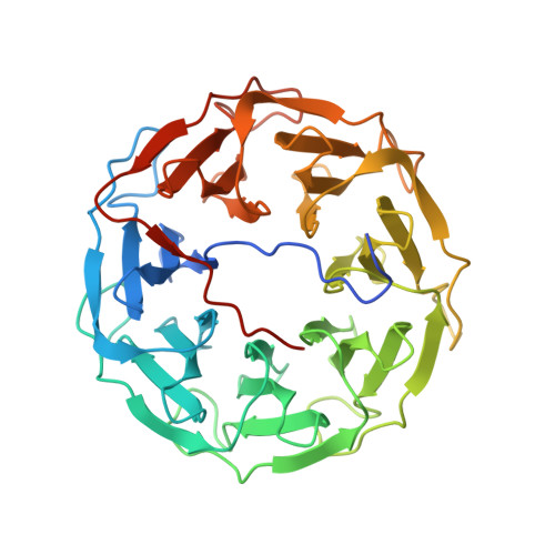
    Photorhabdus luminescens is known for its symbiosis with the entomopathogenic nematode Heterorhabditis bacteriophora and its pathogenicity toward insect larvae. A hypothetical protein from P. luminescens was identified, purified from the native source, and characterized as an l-fucose-binding lectin, named P. luminescens lectin (PLL). Glycan array and biochemical characterization data revealed PLL to be specific toward l-fucose and the disaccharide glycan 3,6-O-Me 2 -Glcβ1-4(2,3-O-Me 2 )Rhaα-O-(p-C 6 H 4 )-OCH 2 CH 2 NH 2 PLL was discovered to be a homotetramer with an intersubunit disulfide bridge. The crystal structures of native and recombinant PLL revealed a seven-bladed β-propeller fold creating seven putative fucose-binding sites per monomer. The crystal structure of the recombinant PLL·l-fucose complex confirmed that at least three sites were fucose-binding. Moreover, the crystal structures indicated that some of the other sites are masked either by the tetrameric nature of the lectin or by incorporation of the C terminus of the lectin into one of these sites. PLL exhibited an ability to bind to insect hemocytes and the cuticular surface of a nematode, H. bacteriophora.


  • Organizational Affiliation

    From the Central European Institute of Technology (CEITEC).


Macromolecules
Find similar proteins by:  (by identity cutoff)  |  3D Structure
Entity ID: 1
MoleculeChains Sequence LengthOrganismDetailsImage
PLL lectin381Photorhabdus luminescensMutation(s): 0 
Gene Names: plu0732
UniProt
Find proteins for Q7N8J0 (Photorhabdus laumondii subsp. laumondii (strain DSM 15139 / CIP 105565 / TT01))
Explore Q7N8J0 
Go to UniProtKB:  Q7N8J0
Entity Groups  
Sequence Clusters30% Identity50% Identity70% Identity90% Identity95% Identity100% Identity
UniProt GroupQ7N8J0
Sequence Annotations
Expand
  • Reference Sequence
Small Molecules
Ligands 2 Unique
IDChains Name / Formula / InChI Key2D Diagram3D Interactions
FUC
Query on FUC

Download Ideal Coordinates CCD File 
B [auth A],
C [auth A],
D [auth A]
alpha-L-fucopyranose
C6 H12 O5
SHZGCJCMOBCMKK-SXUWKVJYSA-N
GOL
Query on GOL

Download Ideal Coordinates CCD File 
E [auth A]
F [auth A]
G [auth A]
H [auth A]
I [auth A]
E [auth A],
F [auth A],
G [auth A],
H [auth A],
I [auth A],
J [auth A],
K [auth A],
L [auth A],
M [auth A],
N [auth A]
GLYCEROL
C3 H8 O3
PEDCQBHIVMGVHV-UHFFFAOYSA-N
Experimental Data & Validation

Experimental Data

  • Method: X-RAY DIFFRACTION
  • Resolution: 1.75 Å
  • R-Value Free: 0.181 
  • R-Value Work: 0.142 
  • R-Value Observed: 0.144 
  • Space Group: I 2 2 2
Unit Cell:
Length ( Å )Angle ( ˚ )
a = 72.783α = 90
b = 87.717β = 90
c = 158.194γ = 90
Software Package:
Software NamePurpose
REFMACrefinement
XDSdata reduction
SCALAdata scaling
MOLREPphasing

Structure Validation

View Full Validation Report


Ligand Structure Quality Assessment 


Entry History & Funding Information

Deposition Data


Funding OrganizationLocationGrant Number
Czech Science FoundationCzech Republic13-25401S
European Regional Development FundCzech RepublicCZ.1.05/1.1.00/02.0068
SYLICACzech RepublicCZ.1.05/1.1.00/02.0068

Revision History  (Full details and data files)

  • Version 1.0: 2016-10-19
    Type: Initial release
  • Version 1.1: 2016-11-02
    Changes: Database references
  • Version 1.2: 2016-12-07
    Changes: Database references
  • Version 1.3: 2020-07-29
    Type: Remediation
    Reason: Carbohydrate remediation
    Changes: Data collection, Derived calculations, Structure summary
  • Version 1.4: 2024-01-10
    Changes: Data collection, Database references, Refinement description, Structure summary