5BYL

Aminoglycoside Phosphotransferase (2'')-Ia (CTD of AAC(6')-Ie/APH(2'')-Ia) in complex with GMPPCP and Magnesium


Experimental Data Snapshot

  • Method: X-RAY DIFFRACTION
  • Resolution: 2.15 Å
  • R-Value Free: 0.198 
  • R-Value Work: 0.156 
  • R-Value Observed: 0.158 

wwPDB Validation   3D Report Full Report


Ligand Structure Quality Assessment 


This is version 1.5 of the entry. See complete history


Literature

Antibiotic Binding Drives Catalytic Activation of Aminoglycoside Kinase APH(2)-Ia.

Caldwell, S.J.Huang, Y.Berghuis, A.M.

(2016) Structure 24: 935-945

  • DOI: https://doi.org/10.1016/j.str.2016.04.002
  • Primary Citation of Related Structures:  
    5BYL, 5IQA, 5IQB, 5IQC, 5IQD, 5IQE, 5IQF, 5IQG, 5IQH, 5IQI

  • PubMed Abstract: 

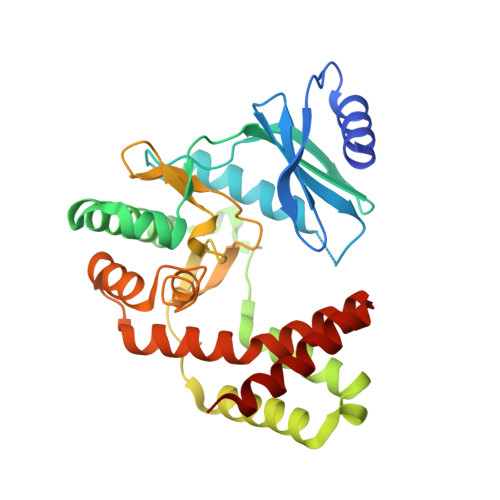
    APH(2″)-Ia is a widely disseminated resistance factor frequently found in clinical isolates of Staphylococcus aureus and pathogenic enterococci, where it is constitutively expressed. APH(2″)-Ia confers high-level resistance to gentamicin and related aminoglycosides through phosphorylation of the antibiotic using guanosine triphosphate (GTP) as phosphate donor. We have determined crystal structures of the APH(2″)-Ia in complex with GTP analogs, guanosine diphosphate, and aminoglycosides. These structures collectively demonstrate that aminoglycoside binding to the GTP-bound kinase drives conformational changes that bring distant regions of the protein into contact. These changes in turn drive a switch of the triphosphate cofactor from an inactive, stabilized conformation to a catalytically competent active conformation. This switch has not been previously reported for antibiotic kinases or for the structurally related eukaryotic protein kinases. This catalytic triphosphate switch presents a means by which the enzyme can curtail wasteful hydrolysis of GTP in the absence of aminoglycosides, providing an evolutionary advantage to this enzyme.


  • Organizational Affiliation

    Department of Biochemistry, McGill University, Montreal, QC H3G 1Y6, Canada; Groupe de Recherche Axé sur la Structure des Protéines, McGill University, Montreal, QC H3G 0B1, Canada.


Macromolecules
Find similar proteins by:  (by identity cutoff)  |  3D Structure
Entity ID: 1
MoleculeChains Sequence LengthOrganismDetailsImage
Bifunctional AAC/APH
A, B, C, D
305Staphylococcus aureusMutation(s): 0 
Gene Names: aacA-aphDR015VRA0030
EC: 2.3.1 (PDB Primary Data), 2.7.1 (PDB Primary Data)
UniProt
Find proteins for P0A0C1 (Staphylococcus aureus)
Explore P0A0C1 
Go to UniProtKB:  P0A0C1
Entity Groups  
Sequence Clusters30% Identity50% Identity70% Identity90% Identity95% Identity100% Identity
UniProt GroupP0A0C1
Sequence Annotations
Expand
  • Reference Sequence
Small Molecules
Ligands 4 Unique
IDChains Name / Formula / InChI Key2D Diagram3D Interactions
GCP
Query on GCP

Download Ideal Coordinates CCD File 
E [auth A],
K [auth B],
P [auth C],
W [auth D]
PHOSPHOMETHYLPHOSPHONIC ACID GUANYLATE ESTER
C11 H18 N5 O13 P3
PHBDHXOBFUBCJD-KQYNXXCUSA-N
GOL
Query on GOL

Download Ideal Coordinates CCD File 
I [auth A]
J [auth A]
O [auth B]
U [auth C]
V [auth C]
I [auth A],
J [auth A],
O [auth B],
U [auth C],
V [auth C],
Z [auth D]
GLYCEROL
C3 H8 O3
PEDCQBHIVMGVHV-UHFFFAOYSA-N
CL
Query on CL

Download Ideal Coordinates CCD File 
H [auth A],
N [auth B],
T [auth C]
CHLORIDE ION
Cl
VEXZGXHMUGYJMC-UHFFFAOYSA-M
MG
Query on MG

Download Ideal Coordinates CCD File 
F [auth A]
G [auth A]
L [auth B]
M [auth B]
Q [auth C]
F [auth A],
G [auth A],
L [auth B],
M [auth B],
Q [auth C],
R [auth C],
S [auth C],
X [auth D],
Y [auth D]
MAGNESIUM ION
Mg
JLVVSXFLKOJNIY-UHFFFAOYSA-N
Experimental Data & Validation

Experimental Data

  • Method: X-RAY DIFFRACTION
  • Resolution: 2.15 Å
  • R-Value Free: 0.198 
  • R-Value Work: 0.156 
  • R-Value Observed: 0.158 
  • Space Group: P 1 21 1
Unit Cell:
Length ( Å )Angle ( ˚ )
a = 90.29α = 90
b = 99.9β = 105.09
c = 93.62γ = 90
Software Package:
Software NamePurpose
REFMACrefinement
MOSFLMdata reduction
Aimlessdata scaling
Cootmodel building
REFMACphasing

Structure Validation

View Full Validation Report


Ligand Structure Quality Assessment 


Entry History & Funding Information

Deposition Data


Funding OrganizationLocationGrant Number
Canadian Institutes of Health Research (CIHR)CanadaMOP-13107

Revision History  (Full details and data files)

  • Version 1.0: 2016-05-25
    Type: Initial release
  • Version 1.1: 2016-06-15
    Changes: Database references
  • Version 1.2: 2017-09-27
    Changes: Author supporting evidence, Database references, Derived calculations
  • Version 1.3: 2017-11-01
    Changes: Author supporting evidence
  • Version 1.4: 2020-01-08
    Changes: Author supporting evidence
  • Version 1.5: 2024-03-06
    Changes: Data collection, Database references, Derived calculations, Refinement description