5BQL

Fluorescent protein cyOFP


Experimental Data Snapshot

  • Method: X-RAY DIFFRACTION
  • Resolution: 2.39 Å
  • R-Value Free: 0.252 
  • R-Value Work: 0.193 
  • R-Value Observed: 0.196 

wwPDB Validation   3D Report Full Report


This is version 1.4 of the entry. See complete history


Literature

A Guide to Fluorescent Protein FRET Pairs.

Bajar, B.T.Wang, E.S.Zhang, S.Lin, M.Z.Chu, J.

(2016) Sensors (Basel) 16

  • DOI: https://doi.org/10.3390/s16091488
  • Primary Citation of Related Structures:  
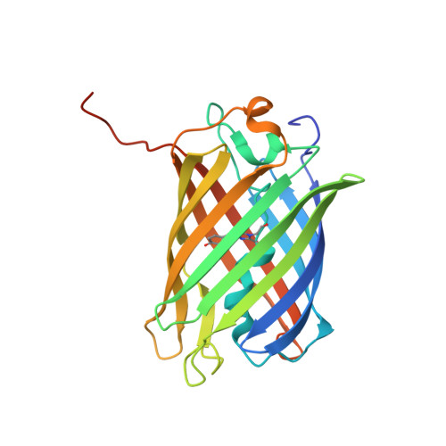
    5BQL

  • PubMed Abstract: 

    Förster or fluorescence resonance energy transfer (FRET) technology and genetically encoded FRET biosensors provide a powerful tool for visualizing signaling molecules in live cells with high spatiotemporal resolution. Fluorescent proteins (FPs) are most commonly used as both donor and acceptor fluorophores in FRET biosensors, especially since FPs are genetically encodable and live-cell compatible. In this review, we will provide an overview of methods to measure FRET changes in biological contexts, discuss the palette of FP FRET pairs developed and their relative strengths and weaknesses, and note important factors to consider when using FPs for FRET studies.


  • Organizational Affiliation

    Medical Scientist Training Program, University of California, Los Angeles, CA 90095, USA. BBajar@mednet.ucla.edu.


Macromolecules
Find similar proteins by:  (by identity cutoff)  |  3D Structure
Entity ID: 1
MoleculeChains Sequence LengthOrganismDetailsImage
Fluorescent protein cyOFP
A, B, C, D
242Entacmaea quadricolorMutation(s): 0 
UniProt
Find proteins for A0A182DW87 (Escherichia coli)
Explore A0A182DW87 
Go to UniProtKB:  A0A182DW87
Entity Groups  
Sequence Clusters30% Identity50% Identity70% Identity90% Identity95% Identity100% Identity
UniProt GroupA0A182DW87
Sequence Annotations
Expand
  • Reference Sequence
Small Molecules
Modified Residues  1 Unique
IDChains TypeFormula2D DiagramParent
NRQ
Query on NRQ
A, B, C, D
L-PEPTIDE LINKINGC16 H17 N3 O4 SMET, TYR, GLY
Experimental Data & Validation

Experimental Data

  • Method: X-RAY DIFFRACTION
  • Resolution: 2.39 Å
  • R-Value Free: 0.252 
  • R-Value Work: 0.193 
  • R-Value Observed: 0.196 
  • Space Group: P 21 21 21
Unit Cell:
Length ( Å )Angle ( ˚ )
a = 74.124α = 90
b = 102.764β = 90
c = 123.254γ = 90
Software Package:
Software NamePurpose
REFMACrefinement
XSCALEdata scaling
PHASERphasing
PDB_EXTRACTdata extraction

Structure Validation

View Full Validation Report


Entry History & Funding Information

Deposition Data


Funding OrganizationLocationGrant Number
National Institutes of Health/National Institute of Neurological Disorders and Stroke (NIH/NINDS)United States1U01NS090600
National Institutes of Health/National Institute of General Medical Sciences (NIH/NIGMS)United StatesP50GM107615

Revision History  (Full details and data files)

  • Version 1.0: 2016-06-01
    Type: Initial release
  • Version 1.1: 2016-10-05
    Changes: Database references
  • Version 1.2: 2017-09-20
    Changes: Author supporting evidence, Derived calculations
  • Version 1.3: 2018-10-10
    Changes: Data collection, Source and taxonomy, Structure summary
  • Version 1.4: 2019-12-18
    Changes: Author supporting evidence, Structure summary