5B6S

Catalytic domain of Coprinopsis cinerea GH62 alpha-L-arabinofuranosidase


Experimental Data Snapshot

  • Method: X-RAY DIFFRACTION
  • Resolution: 1.70 Å
  • R-Value Free: 0.170 
  • R-Value Work: 0.139 
  • R-Value Observed: 0.141 

wwPDB Validation   3D Report Full Report


This is version 1.4 of the entry. See complete history


Literature

Structure of the Catalytic Domain of alpha-L-Arabinofuranosidase from Coprinopsis cinerea, CcAbf62A, Provides Insights into Structure-Function Relationships in Glycoside Hydrolase Family 62

Tonozuka, T.Tanaka, Y.Okuyama, S.Miyazaki, T.Nishikawa, A.Yoshida, M.

(2017) Appl Biochem Biotechnol 181: 511-525

  • DOI: https://doi.org/10.1007/s12010-016-2227-0
  • Primary Citation of Related Structures:  
    5B6S, 5B6T

  • PubMed Abstract: 

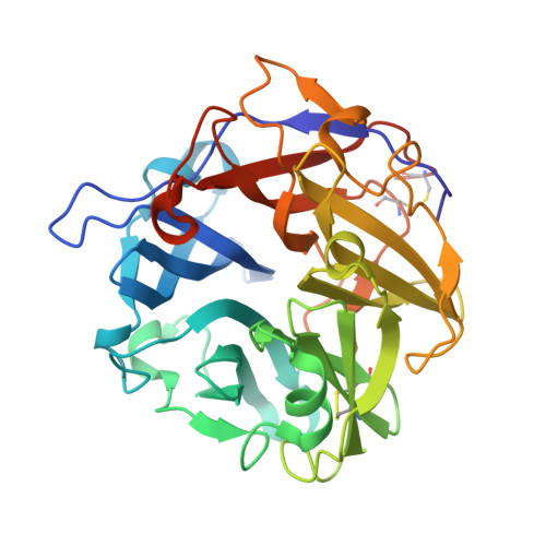
    α-L-Arabinofuranosidases, belonging to the glycoside hydrolase family (GH) 62, hydrolyze the α-1,2- or α-1,3-bond to liberate L-arabinofuranose from the xylan backbone. Here, we determined the structure of the C-terminal catalytic domain of CcAbf62A, a GH62 α-L-arabinofuranosidase from Coprinopsis cinerea. CcAbf62A is composed of a five-bladed β-propeller, as observed in other GH62 enzymes. The structure near the active site of CcAbf62A is also highly homologous to those of other GH62 enzymes. However, a calcium atom in the catalytic center interacts with an asparagine residue, Asn279, which is not found in other GH62 enzymes. In addition, some residues in subsites +3R, +2NR, +3NR, and +4NR of CcAbf62A are not conserved in other GH62 enzymes. In particular, a histidine residue, His221, is uniquely observed in subsite +2NR of CcAbf62A, which is likely to influence the substrate specificity. The results obtained here suggest that the amino acid residues that interact with the xylan backbone vary among the GH62 enzymes, despite the high similarity of their overall structures.


  • Organizational Affiliation

    Department of Applied Biological Science, Tokyo University of Agriculture and Technology, Tokyo, 183-8509, Japan. tonozuka@cc.tuat.ac.jp.


Macromolecules
Find similar proteins by:  (by identity cutoff)  |  3D Structure
Entity ID: 1
MoleculeChains Sequence LengthOrganismDetailsImage
Glycosyl hydrolase family 62 protein
A, B
327Coprinopsis cinerea okayama7#130Mutation(s): 0 
Gene Names: CC1G_01577
EC: 3.2.1.55
UniProt
Find proteins for A8NI40 (Coprinopsis cinerea (strain Okayama-7 / 130 / ATCC MYA-4618 / FGSC 9003))
Explore A8NI40 
Go to UniProtKB:  A8NI40
Entity Groups  
Sequence Clusters30% Identity50% Identity70% Identity90% Identity95% Identity100% Identity
UniProt GroupA8NI40
Sequence Annotations
Expand
  • Reference Sequence
Experimental Data & Validation

Experimental Data

  • Method: X-RAY DIFFRACTION
  • Resolution: 1.70 Å
  • R-Value Free: 0.170 
  • R-Value Work: 0.139 
  • R-Value Observed: 0.141 
  • Space Group: P 1 21 1
Unit Cell:
Length ( Å )Angle ( ˚ )
a = 47.713α = 90
b = 77.735β = 93
c = 74.202γ = 90
Software Package:
Software NamePurpose
REFMACrefinement
HKL-2000data reduction
HKL-2000data scaling
MOLREPphasing

Structure Validation

View Full Validation Report


Entry History & Funding Information

Deposition Data

  • Released Date: 2016-09-07 
  • Deposition Author(s): Tonozuka, T.

Funding OrganizationLocationGrant Number
Japan Society for the Promotion of ScienceJapan16K07687

Revision History  (Full details and data files)

  • Version 1.0: 2016-09-07
    Type: Initial release
  • Version 1.1: 2016-09-14
    Changes: Database references
  • Version 1.2: 2017-02-15
    Changes: Database references
  • Version 1.3: 2020-02-26
    Changes: Data collection
  • Version 1.4: 2023-11-08
    Changes: Data collection, Database references, Derived calculations, Refinement description