5B5R

Crystal structure of GSDMA3


Experimental Data Snapshot

  • Method: X-RAY DIFFRACTION
  • Resolution: 1.90 Å
  • R-Value Free: 0.232 
  • R-Value Work: 0.187 
  • R-Value Observed: 0.189 

wwPDB Validation   3D Report Full Report


This is version 1.2 of the entry. See complete history


Literature

Pore-forming activity and structural autoinhibition of the gasdermin family.

Ding, J.Wang, K.Liu, W.She, Y.Sun, Q.Shi, J.Sun, H.Wang, D.C.Shao, F.

(2016) Nature 535: 111-116

  • DOI: https://doi.org/10.1038/nature18590
  • Primary Citation of Related Structures:  
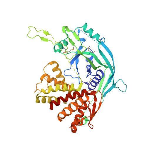
    5B5R

  • PubMed Abstract: 

    Inflammatory caspases cleave the gasdermin D (GSDMD) protein to trigger pyroptosis, a lytic form of cell death that is crucial for immune defences and diseases. GSDMD contains a functionally important gasdermin-N domain that is shared in the gasdermin family. The functional mechanism of action of gasdermin proteins is unknown. Here we show that the gasdermin-N domains of the gasdermin proteins GSDMD, GSDMA3 and GSDMA can bind membrane lipids, phosphoinositides and cardiolipin, and exhibit membrane-disrupting cytotoxicity in mammalian cells and artificially transformed bacteria. Gasdermin-N moved to the plasma membrane during pyroptosis. Purified gasdermin-N efficiently lysed phosphoinositide/cardiolipin-containing liposomes and formed pores on membranes made of artificial or natural phospholipid mixtures. Most gasdermin pores had an inner diameter of 10–14 nm and contained 16 symmetric protomers. The crystal structure of GSDMA3 showed an autoinhibited two-domain architecture that is conserved in the gasdermin family. Structure-guided mutagenesis demonstrated that the liposome-leakage and pore-forming activities of the gasdermin-N domain are required for pyroptosis. These findings reveal the mechanism for pyroptosis and provide insights into the roles of the gasdermin family in necrosis, immunity and diseases.


Macromolecules
Find similar proteins by:  (by identity cutoff)  |  3D Structure
Entity ID: 1
MoleculeChains Sequence LengthOrganismDetailsImage
Gasdermin-A3468Mus musculusMutation(s): 0 
Gene Names: Gsdma3Gsdm3
Membrane Entity: Yes 
UniProt & NIH Common Fund Data Resources
Find proteins for Q5Y4Y6 (Mus musculus)
Explore Q5Y4Y6 
Go to UniProtKB:  Q5Y4Y6
IMPC:  MGI:3044668
Entity Groups  
Sequence Clusters30% Identity50% Identity70% Identity90% Identity95% Identity100% Identity
UniProt GroupQ5Y4Y6
Sequence Annotations
Expand
  • Reference Sequence
Small Molecules
Modified Residues  1 Unique
IDChains TypeFormula2D DiagramParent
MSE
Query on MSE
A
L-PEPTIDE LINKINGC5 H11 N O2 SeMET
Experimental Data & Validation

Experimental Data

  • Method: X-RAY DIFFRACTION
  • Resolution: 1.90 Å
  • R-Value Free: 0.232 
  • R-Value Work: 0.187 
  • R-Value Observed: 0.189 
  • Space Group: P 1 21 1
Unit Cell:
Length ( Å )Angle ( ˚ )
a = 43.453α = 90
b = 103.414β = 110.57
c = 49.625γ = 90
Software Package:
Software NamePurpose
PHENIXrefinement
HKL-2000data reduction
SCALEPACKdata scaling
PHENIXphasing

Structure Validation

View Full Validation Report


Entry History 

Deposition Data

Revision History  (Full details and data files)

  • Version 1.0: 2016-06-15
    Type: Initial release
  • Version 1.1: 2016-07-20
    Changes: Database references
  • Version 1.2: 2017-09-27
    Changes: Data collection, Database references, Derived calculations