5B5Q

1.7 Angstroms structure of ChlaDub1 from Chlamydia Trachomatis


Experimental Data Snapshot

  • Method: X-RAY DIFFRACTION
  • Resolution: 1.70 Å
  • R-Value Free: 0.213 
  • R-Value Work: 0.165 
  • R-Value Observed: 0.168 

wwPDB Validation   3D Report Full Report


This is version 1.1 of the entry. See complete history


Literature

Chlamydia trachomatis-containing vacuole serves as deubiquitination platform to stabilize Mcl-1 and to interfere with host defense

Fischer, A.Harrison, K.S.Ramirez, Y.Auer, D.Chowdhury, S.R.Prusty, B.K.Sauer, F.Dimond, Z.Kisker, C.Scott Hefty, P.Rudel, T.

(2017) Elife 6

  • DOI: https://doi.org/10.7554/eLife.21465
  • Primary Citation of Related Structures:  
    5B5Q

  • PubMed Abstract: 

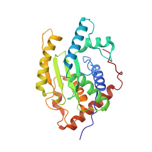
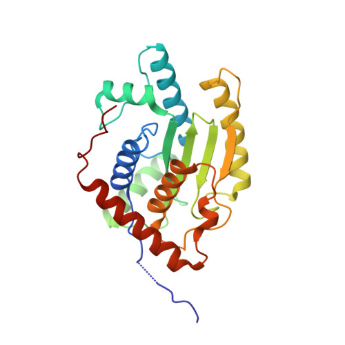
    Obligate intracellular Chlamydia trachomatis replicate in a membrane-bound vacuole called inclusion, which serves as a signaling interface with the host cell. Here, we show that the chlamydial deubiquitinating enzyme (Cdu) 1 localizes in the inclusion membrane and faces the cytosol with the active deubiquitinating enzyme domain. The structure of this domain revealed high similarity to mammalian deubiquitinases with a unique α-helix close to the substrate-binding pocket. We identified the apoptosis regulator Mcl-1 as a target that interacts with Cdu1 and is stabilized by deubiquitination at the chlamydial inclusion. A chlamydial transposon insertion mutant in the Cdu1-encoding gene exhibited increased Mcl-1 and inclusion ubiquitination and reduced Mcl-1 stabilization. Additionally, inactivation of Cdu1 led to increased sensitivity of C. trachomatis for IFNγ and impaired infection in mice. Thus, the chlamydial inclusion serves as an enriched site for a deubiquitinating activity exerting a function in selective stabilization of host proteins and protection from host defense.


  • Organizational Affiliation

    Department of Microbiology, Biocenter, University of Würzburg, Würzburg, Germany.


Macromolecules
Find similar proteins by:  (by identity cutoff)  |  3D Structure
Entity ID: 1
MoleculeChains Sequence LengthOrganismDetailsImage
Membrane thiol protease243Chlamydia trachomatisMutation(s): 0 
Gene Names: cdu1ERS066953_00737
EC: 3.4.22
UniProt
Find proteins for B0B9A0 (Chlamydia trachomatis serovar L2 (strain 434/Bu / ATCC VR-902B))
Explore B0B9A0 
Go to UniProtKB:  B0B9A0
Entity Groups  
Sequence Clusters30% Identity50% Identity70% Identity90% Identity95% Identity100% Identity
UniProt GroupB0B9A0
Sequence Annotations
Expand
  • Reference Sequence
Find similar proteins by:  (by identity cutoff)  |  3D Structure
Entity ID: 2
MoleculeChains Sequence LengthOrganismDetailsImage
Membrane thiol protease249Chlamydia trachomatisMutation(s): 0 
Gene Names: CT868
UniProt
Find proteins for B0B9A0 (Chlamydia trachomatis serovar L2 (strain 434/Bu / ATCC VR-902B))
Explore B0B9A0 
Go to UniProtKB:  B0B9A0
Entity Groups  
Sequence Clusters30% Identity50% Identity70% Identity90% Identity95% Identity100% Identity
UniProt GroupB0B9A0
Sequence Annotations
Expand
  • Reference Sequence
Small Molecules
Ligands 1 Unique
IDChains Name / Formula / InChI Key2D Diagram3D Interactions
CL
Query on CL

Download Ideal Coordinates CCD File 
C [auth B]CHLORIDE ION
Cl
VEXZGXHMUGYJMC-UHFFFAOYSA-M
Experimental Data & Validation

Experimental Data

  • Method: X-RAY DIFFRACTION
  • Resolution: 1.70 Å
  • R-Value Free: 0.213 
  • R-Value Work: 0.165 
  • R-Value Observed: 0.168 
  • Space Group: P 1 21 1
Unit Cell:
Length ( Å )Angle ( ˚ )
a = 39.1α = 90
b = 77.573β = 96.43
c = 68.948γ = 90
Software Package:
Software NamePurpose
PHENIXrefinement
XDSdata reduction
Aimlessdata scaling
PHASERphasing

Structure Validation

View Full Validation Report


Entry History & Funding Information

Deposition Data


Funding OrganizationLocationGrant Number
GSLS University of WuerzburgGermany--

Revision History  (Full details and data files)

  • Version 1.0: 2017-03-29
    Type: Initial release
  • Version 1.1: 2017-11-22
    Changes: Database references