5AWM

The Crystal Structure of JNK from Drosophila melanogaster Reveals an Evolutionarily Conserved Topology with that of Mammalian JNK Proteins.


Experimental Data Snapshot

  • Method: X-RAY DIFFRACTION
  • Resolution: 1.79 Å
  • R-Value Free: 0.220 
  • R-Value Work: 0.175 
  • R-Value Observed: 0.179 

wwPDB Validation   3D Report Full Report


Ligand Structure Quality Assessment 


This is version 1.3 of the entry. See complete history

Re-refinement Note

This entry reflects an alternative modeling of the original data in: 4M3A


Literature

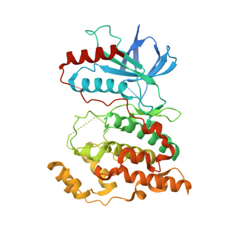
The crystal structure of JNK from Drosophila melanogaster reveals an evolutionarily conserved topology with that of mammalian JNK proteins.

Chimnaronk, S.Sitthiroongruang, J.Srisucharitpanit, K.Srisaisup, M.Ketterman, A.J.Boonserm, P.

(2015) BMC Struct Biol 15: 17-17

  • DOI: https://doi.org/10.1186/s12900-015-0045-1
  • Primary Citation of Related Structures:  
    5AWM

  • PubMed Abstract: 

    The c-Jun N-terminal kinases (JNKs), members of the mitogen-activated protein kinase (MAPK) family, engage in diverse cellular responses to signals produced under normal development and stress conditions. In Drosophila, only one JNK member is present, whereas ten isoforms from three JNK genes (JNK1, 2, and 3) are present in mammalian cells. To date, several mammalian JNK structures have been determined, however, there has been no report of any insect JNK structure.


  • Organizational Affiliation

    Institute of Molecular Biosciences, Mahidol University, Salaya, Phuttamonthon, Nakhon Pathom, 73170, Thailand. sarin.chi@mahidol.ac.th.


Macromolecules
Find similar proteins by:  (by identity cutoff)  |  3D Structure
Entity ID: 1
MoleculeChains Sequence LengthOrganismDetailsImage
Stress-activated protein kinase JNK394Drosophila melanogasterMutation(s): 0 
Gene Names: bskJNKCG5680
EC: 2.7.11.24
UniProt
Find proteins for P92208 (Drosophila melanogaster)
Explore P92208 
Go to UniProtKB:  P92208
Entity Groups  
Sequence Clusters30% Identity50% Identity70% Identity90% Identity95% Identity100% Identity
UniProt GroupP92208
Sequence Annotations
Expand
  • Reference Sequence
Experimental Data & Validation

Experimental Data

  • Method: X-RAY DIFFRACTION
  • Resolution: 1.79 Å
  • R-Value Free: 0.220 
  • R-Value Work: 0.175 
  • R-Value Observed: 0.179 
  • Space Group: P 21 21 21
Unit Cell:
Length ( Å )Angle ( ˚ )
a = 52.486α = 90
b = 55.329β = 90
c = 126.717γ = 90
Software Package:
Software NamePurpose
REFMACrefinement
XDSdata reduction
HKL-2000data scaling
MOLREPphasing

Structure Validation

View Full Validation Report


Ligand Structure Quality Assessment 


Entry History & Funding Information

Deposition Data

  • Released Date: 2015-08-05 
  • Deposition Author(s): Boonserm, P.
  • This entry supersedes: 4M3A

Funding OrganizationLocationGrant Number
Mahidol UniversityThailand--

Revision History  (Full details and data files)

  • Version 1.0: 2015-08-05
    Type: Initial release
  • Version 1.1: 2015-11-11
    Changes: Database references
  • Version 1.2: 2020-02-26
    Changes: Data collection, Derived calculations
  • Version 1.3: 2023-11-08
    Changes: Data collection, Database references, Derived calculations, Refinement description