5A8V

Crystal structure of Orgyia pseudotsugata CPV5 polyhedra with SeMet substitution


Experimental Data Snapshot

  • Method: X-RAY DIFFRACTION
  • Resolution: 2.07 Å
  • R-Value Free: 0.228 
  • R-Value Work: 0.169 
  • R-Value Observed: 0.172 

wwPDB Validation   3D Report Full Report


This is version 1.2 of the entry. See complete history


Literature

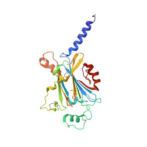
Polyhedra Structures and the Evolution of the Insect Viruses.

Ji, X.Axford, D.Owen, R.Evans, G.Ginn, H.M.Sutton, G.Stuart, D.I.

(2015) J Struct Biol 192: 88

  • DOI: https://doi.org/10.1016/j.jsb.2015.08.009
  • Primary Citation of Related Structures:  
    5A8S, 5A8T, 5A8U, 5A8V, 5A96, 5A98, 5A99, 5A9A, 5A9B, 5A9C, 5A9P

  • PubMed Abstract: 

    Polyhedra represent an ancient system used by a number of insect viruses to protect virions during long periods of environmental exposure. We present high resolution crystal structures of polyhedra for seven previously uncharacterised types of cypoviruses, four using ab initio selenomethionine phasing (two of these required over 100 selenomethionine crystals each). Approximately 80% of residues are structurally equivalent between all polyhedrins (pairwise rmsd ⩽ 1.5 Å), whilst pairwise sequence identities, based on structural alignment, are as little as 12%. These structures illustrate the effect of 400 million years of evolution on a system where the crystal lattice is the functionally conserved feature in the face of massive sequence variability. The conservation of crystal contacts is maintained across most of the molecular surface, except for a dispensable virus recognition domain. By spreading the contacts over so much of the protein surface the lattice remains robust in the face of many individual changes. Overall these unusual structural constraints seem to have skewed the molecule's evolution so that surface residues are almost as conserved as the internal residues.


  • Organizational Affiliation

    Division of Structural Biology, The Wellcome Trust Centre for Human Genetics, University of Oxford, Roosevelt Drive, Oxford, Oxfordshire OX3 7BN, United Kingdom.


Macromolecules
Find similar proteins by:  (by identity cutoff)  |  3D Structure
Entity ID: 1
MoleculeChains Sequence LengthOrganismDetailsImage
POLYHEDRIN253Orgyia pseudotsugata cypovirus 5Mutation(s): 1 
UniProt
Find proteins for P36701 (Orgyia pseudotsugata cypovirus)
Explore P36701 
Go to UniProtKB:  P36701
Entity Groups  
Sequence Clusters30% Identity50% Identity70% Identity90% Identity95% Identity100% Identity
UniProt GroupP36701
Sequence Annotations
Expand
  • Reference Sequence
Small Molecules
Ligands 1 Unique
IDChains Name / Formula / InChI Key2D Diagram3D Interactions
CA
Query on CA

Download Ideal Coordinates CCD File 
B [auth A]CALCIUM ION
Ca
BHPQYMZQTOCNFJ-UHFFFAOYSA-N
Modified Residues  1 Unique
IDChains TypeFormula2D DiagramParent
MSE
Query on MSE
A
L-PEPTIDE LINKINGC5 H11 N O2 SeMET
Experimental Data & Validation

Experimental Data

  • Method: X-RAY DIFFRACTION
  • Resolution: 2.07 Å
  • R-Value Free: 0.228 
  • R-Value Work: 0.169 
  • R-Value Observed: 0.172 
  • Space Group: I 2 3
Unit Cell:
Length ( Å )Angle ( ˚ )
a = 102.054α = 90
b = 102.054β = 90
c = 102.054γ = 90
Software Package:
Software NamePurpose
PHENIXrefinement
xia2data reduction
xia2data scaling
SHARPphasing

Structure Validation

View Full Validation Report


Entry History 

Deposition Data

Revision History  (Full details and data files)

  • Version 1.0: 2015-09-02
    Type: Initial release
  • Version 1.1: 2015-10-14
    Changes: Database references
  • Version 1.2: 2019-10-23
    Changes: Data collection, Database references, Derived calculations, Other