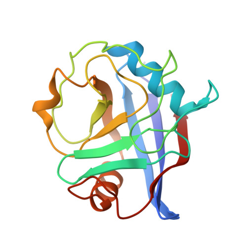
4ZSC

Human Cyclophilin D Complexed with an Inhibitor at room temperature


Experimental Data Snapshot

  • Method: X-RAY DIFFRACTION
  • Resolution: 1.50 Å
  • R-Value Free: 0.163 
  • R-Value Work: 0.134 
  • R-Value Observed: 0.135 

wwPDB Validation   3D Report Full Report


Ligand Structure Quality Assessment 


This is version 1.1 of the entry. See complete history


Literature

Combining `dry' co-crystallization and in situ diffraction to facilitate ligand screening by X-ray crystallography.

Gelin, M.Delfosse, V.Allemand, F.Hoh, F.Sallaz-Damaz, Y.Pirocchi, M.Bourguet, W.Ferrer, J.L.Labesse, G.Guichou, J.F.

(2015) Acta Crystallogr D Biol Crystallogr 71: 1777-1787

  • DOI: https://doi.org/10.1107/S1399004715010342
  • Primary Citation of Related Structures:  
    3RDC, 4XLD, 4XN6, 4XNC, 4XNE, 4XOY, 4XOZ, 4XP0, 4XP2, 4XP3, 4XRJ, 4XRL, 4ZSC, 4ZSD

  • PubMed Abstract: 

    X-ray crystallography is an established technique for ligand screening in fragment-based drug-design projects, but the required manual handling steps - soaking crystals with ligand and the subsequent harvesting - are tedious and limit the throughput of the process. Here, an alternative approach is reported: crystallization plates are pre-coated with potential binders prior to protein crystallization and X-ray diffraction is performed directly 'in situ' (or in-plate). Its performance is demonstrated on distinct and relevant therapeutic targets currently being studied for ligand screening by X-ray crystallography using either a bending-magnet beamline or a rotating-anode generator. The possibility of using DMSO stock solutions of the ligands to be coated opens up a route to screening most chemical libraries.


  • Organizational Affiliation

    CNRS, UMR5048 - Université de Montpellier, Centre de Biochimie Structurale, 34090 Montpellier, France.


Macromolecules
Find similar proteins by:  (by identity cutoff)  |  3D Structure
Entity ID: 1
MoleculeChains Sequence LengthOrganismDetailsImage
Peptidyl-prolyl cis-trans isomerase F, mitochondrial164Homo sapiensMutation(s): 1 
Gene Names: PPIFCYP3
EC: 5.2.1.8
UniProt & NIH Common Fund Data Resources
Find proteins for P30405 (Homo sapiens)
Explore P30405 
Go to UniProtKB:  P30405
PHAROS:  P30405
GTEx:  ENSG00000108179 
Entity Groups  
Sequence Clusters30% Identity50% Identity70% Identity90% Identity95% Identity100% Identity
UniProt GroupP30405
Sequence Annotations
Expand
  • Reference Sequence
Small Molecules
Ligands 1 Unique
IDChains Name / Formula / InChI Key2D Diagram3D Interactions
EA4
Query on EA4

Download Ideal Coordinates CCD File 
B [auth A]ethyl N-[(4-aminobenzyl)carbamoyl]glycinate
C12 H17 N3 O3
UCMPTJMHPJQEED-UHFFFAOYSA-N
Experimental Data & Validation

Experimental Data

  • Method: X-RAY DIFFRACTION
  • Resolution: 1.50 Å
  • R-Value Free: 0.163 
  • R-Value Work: 0.134 
  • R-Value Observed: 0.135 
  • Space Group: P 41 21 2
Unit Cell:
Length ( Å )Angle ( ˚ )
a = 57.866α = 90
b = 57.866β = 90
c = 88.463γ = 90
Software Package:
Software NamePurpose
REFMACrefinement
SCALAdata scaling
PDB_EXTRACTdata extraction
XSCALEdata scaling
Cootmodel building
XDSdata reduction
REFMACphasing

Structure Validation

View Full Validation Report


Ligand Structure Quality Assessment 


Entry History 

Deposition Data

Revision History  (Full details and data files)

  • Version 1.0: 2015-08-12
    Type: Initial release
  • Version 1.1: 2015-08-19
    Changes: Database references