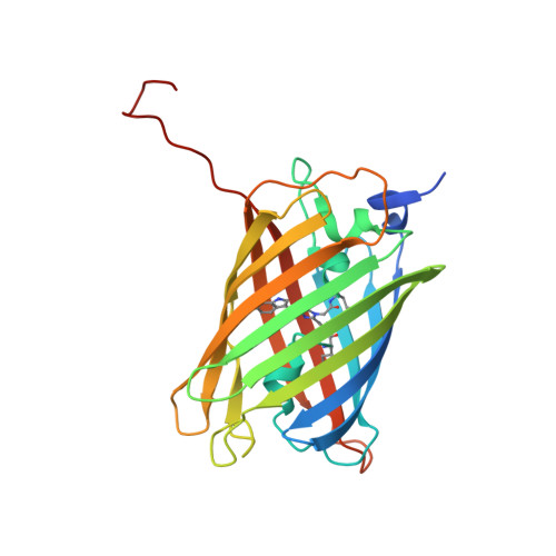
4ZFS

Phototoxic Fluorescent Protein KillerOrange


Experimental Data Snapshot

  • Method: X-RAY DIFFRACTION
  • Resolution: 2.01 Å
  • R-Value Free: 0.236 
  • R-Value Work: 0.192 
  • R-Value Observed: 0.192 

wwPDB Validation   3D Report Full Report


This is version 2.1 of the entry. See complete history


Literature

Crystal Structure of Phototoxic Orange Fluorescent Proteins with a Tryptophan-Based Chromophore.

Pletneva, N.V.Pletnev, V.Z.Sarkisyan, K.S.Gorbachev, D.A.Egorov, E.S.Mishin, A.S.Lukyanov, K.A.Dauter, Z.Pletnev, S.

(2015) PLoS One 10: e0145740-e0145740

  • DOI: https://doi.org/10.1371/journal.pone.0145740
  • Primary Citation of Related Structures:  
    4ZBL, 4ZFS

  • PubMed Abstract: 

    Phototoxic fluorescent proteins represent a sparse group of genetically encoded photosensitizers that could be used for precise light-induced inactivation of target proteins, DNA damage, and cell killing. Only two such GFP-based fluorescent proteins (FPs), KillerRed and its monomeric variant SuperNova, were described up to date. Here, we present a crystallographic study of their two orange successors, dimeric KillerOrange and monomeric mKillerOrange, at 1.81 and 1.57 Å resolution, respectively. They are the first orange-emitting protein photosensitizers with a tryptophan-based chromophore (Gln65-Trp66-Gly67). Same as their red progenitors, both orange photosensitizers have a water-filled channel connecting the chromophore to the β-barrel exterior and enabling transport of ROS. In both proteins, Trp66 of the chromophore adopts an unusual trans-cis conformation stabilized by H-bond with the nearby Gln159. This trans-cis conformation along with the water channel was shown to be a key structural feature providing bright orange emission and phototoxicity of both examined orange photosensitizers.


  • Organizational Affiliation

    Shemyakin-Ovchinnikov Institute of Bioorganic Chemistry, Russian Academy of Sciences, Moscow, Russian Federation.


Macromolecules
Find similar proteins by:  (by identity cutoff)  |  3D Structure
Entity ID: 1
MoleculeChains Sequence LengthOrganismDetailsImage
KillerOrange
A, B, C, D, E
246HydrozoaMutation(s): 0 
UniProt
Find proteins for Q2TCH5 (Anthomedusae sp. DC-2005)
Explore Q2TCH5 
Go to UniProtKB:  Q2TCH5
Entity Groups  
Sequence Clusters30% Identity50% Identity70% Identity90% Identity95% Identity100% Identity
UniProt GroupQ2TCH5
Sequence Annotations
Expand
  • Reference Sequence
Experimental Data & Validation

Experimental Data

  • Method: X-RAY DIFFRACTION
  • Resolution: 2.01 Å
  • R-Value Free: 0.236 
  • R-Value Work: 0.192 
  • R-Value Observed: 0.192 
  • Space Group: C 2 2 21
Unit Cell:
Length ( Å )Angle ( ˚ )
a = 128.93α = 90
b = 202.064β = 90
c = 116.679γ = 90
Software Package:
Software NamePurpose
HKL-2000data reduction
SCALEPACKdata scaling
MOLREPphasing
REFMACrefinement
PDB_EXTRACTdata extraction

Structure Validation

View Full Validation Report


Entry History & Funding Information

Deposition Data


Funding OrganizationLocationGrant Number
Russian Science FoundationRussian Federation14-14-00281

Revision History  (Full details and data files)

  • Version 1.0: 2015-12-23
    Type: Initial release
  • Version 1.1: 2016-01-13
    Changes: Database references
  • Version 2.0: 2023-11-15
    Changes: Atomic model, Data collection, Database references, Derived calculations
  • Version 2.1: 2024-01-10
    Changes: Refinement description