


Crystal structure of S-hydroxynitrile lyase from Manihot esculenta (His103Leu)
Dadashipour, M., Nakano, S., Asano, Y.To be published.
Experimental Data Snapshot
wwPDB Validation 3D Report Full Report

Entity ID: 1 | |||||
|---|---|---|---|---|---|
| Molecule | Chains | Sequence Length | Organism | Details | Image |
| (S)-hydroxynitrile lyase | 258 | Manihot esculenta | Mutation(s): 1 Gene Names: HNL EC: 4.1.2.47 | 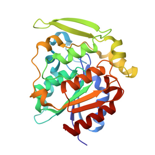 | |
UniProt | |||||
Find proteins for P52705 (Manihot esculenta) Explore P52705 Go to UniProtKB: P52705 | |||||
Entity Groups | |||||
| Sequence Clusters | 30% Identity50% Identity70% Identity90% Identity95% Identity100% Identity | ||||
| UniProt Group | P52705 | ||||
Sequence AnnotationsExpand | |||||
| |||||
| Length ( Å ) | Angle ( ˚ ) |
|---|---|
| a = 86.469 | α = 90 |
| b = 88.098 | β = 90 |
| c = 135.213 | γ = 90 |
| Software Name | Purpose |
|---|---|
| REFMAC | refinement |
| HKL-2000 | data reduction |
| HKL-2000 | data scaling |
| MOLREP | phasing |
RCSB PDB (citation) is hosted by
RCSB PDB is a member of the

