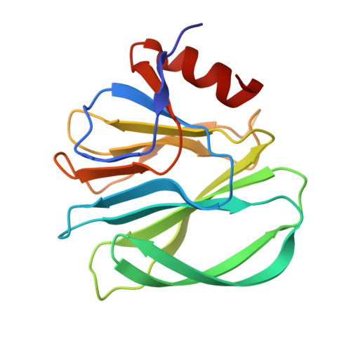
4YG3

Structural basis of glycan recognition in neonate-specific rotaviruses


Experimental Data Snapshot

  • Method: X-RAY DIFFRACTION
  • Resolution: 2.29 Å
  • R-Value Free: 0.242 
  • R-Value Work: 0.184 
  • R-Value Observed: 0.187 

wwPDB Validation   3D Report Full Report


This is version 1.5 of the entry. See complete history


Literature

Structural basis of glycan specificity in neonate-specific bovine-human reassortant rotavirus.

Hu, L.Ramani, S.Czako, R.Sankaran, B.Yu, Y.Smith, D.F.Cummings, R.D.Estes, M.K.Venkataram Prasad, B.V.

(2015) Nat Commun 6: 8346-8346

  • DOI: https://doi.org/10.1038/ncomms9346
  • Primary Citation of Related Structures:  
    4YFW, 4YFZ, 4YG0, 4YG3, 4YG6

  • PubMed Abstract: 

    Strain-dependent variation of glycan recognition during initial cell attachment of viruses is a critical determinant of host specificity, tissue-tropism and zoonosis. Rotaviruses (RVs), which cause life-threatening gastroenteritis in infants and children, display significant genotype-dependent variations in glycan recognition resulting from sequence alterations in the VP8* domain of the spike protein VP4. The structural basis of this genotype-dependent glycan specificity, particularly in human RVs, remains poorly understood. Here, from crystallographic studies, we show how genotypic variations configure a novel binding site in the VP8* of a neonate-specific bovine-human reassortant to uniquely recognize either type I or type II precursor glycans, and to restrict type II glycan binding in the bovine counterpart. Such a distinct glycan-binding site that allows differential recognition of the precursor glycans, which are developmentally regulated in the neonate gut and abundant in bovine and human milk provides a basis for age-restricted tropism and zoonotic transmission of G10P[11] rotaviruses.


  • Organizational Affiliation

    Verna and Marrs McLean Department of Biochemistry and Molecular Biology, Baylor College of Medicine, One Baylor Plaza, Houston, Texas 77030, USA.


Macromolecules
Find similar proteins by:  (by identity cutoff)  |  3D Structure
Entity ID: 1
MoleculeChains Sequence LengthOrganismDetailsImage
Outer capsid protein VP4163Rotavirus A RVA/Cow-tc/USA/B223/1983/G10P[11]Mutation(s): 0 
UniProt
Find proteins for A2T3T2 (Rotavirus A (isolate RVA/Monkey/South Africa/SA11-H96/1958/G3P5B[2]))
Explore A2T3T2 
Go to UniProtKB:  A2T3T2
Entity Groups  
Sequence Clusters30% Identity50% Identity70% Identity90% Identity95% Identity100% Identity
UniProt GroupA2T3T2
Sequence Annotations
Expand
  • Reference Sequence
Experimental Data & Validation

Experimental Data

  • Method: X-RAY DIFFRACTION
  • Resolution: 2.29 Å
  • R-Value Free: 0.242 
  • R-Value Work: 0.184 
  • R-Value Observed: 0.187 
  • Space Group: P 1 21 1
Unit Cell:
Length ( Å )Angle ( ˚ )
a = 28.812α = 90
b = 64.734β = 114.87
c = 36.752γ = 90
Software Package:
Software NamePurpose
PHENIXrefinement
HKL-2000data reduction
MOSFLMdata reduction
PHASERphasing

Structure Validation

View Full Validation Report


Entry History & Funding Information

Deposition Data


Funding OrganizationLocationGrant Number
National Institutes of Health/National Institute Of Allergy and Infectious Diseases (NIH/NIAID)United StatesAI36040

Revision History  (Full details and data files)

  • Version 1.0: 2015-09-30
    Type: Initial release
  • Version 1.1: 2015-10-14
    Changes: Database references
  • Version 1.2: 2017-09-13
    Changes: Author supporting evidence, Derived calculations
  • Version 1.3: 2017-11-22
    Changes: Refinement description
  • Version 1.4: 2019-12-11
    Changes: Author supporting evidence
  • Version 1.5: 2023-09-27
    Changes: Data collection, Database references, Refinement description