4YCO

E. coli dihydrouridine synthase C (DusC) in complex with tRNAPhe


Experimental Data Snapshot

  • Method: X-RAY DIFFRACTION
  • Resolution: 2.10 Å
  • R-Value Free: 0.223 
  • R-Value Work: 0.192 
  • R-Value Observed: 0.193 

wwPDB Validation   3D Report Full Report


Ligand Structure Quality Assessment 


This is version 1.3 of the entry. See complete history


Literature

Major reorientation of tRNA substrates defines specificity of dihydrouridine synthases.

Byrne, R.T.Jenkins, H.T.Peters, D.T.Whelan, F.Stowell, J.Aziz, N.Kasatsky, P.Rodnina, M.V.Koonin, E.V.Konevega, A.L.Antson, A.A.

(2015) Proc Natl Acad Sci U S A 112: 6033-6037

  • DOI: https://doi.org/10.1073/pnas.1500161112
  • Primary Citation of Related Structures:  
    4BF9, 4BFA, 4YCO, 4YCP

  • PubMed Abstract: 

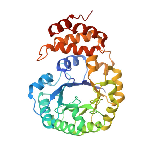
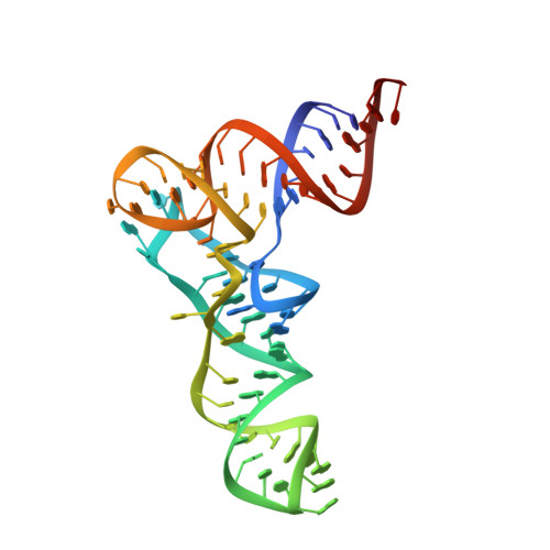
    The reduction of specific uridines to dihydrouridine is one of the most common modifications in tRNA. Increased levels of the dihydrouridine modification are associated with cancer. Dihydrouridine synthases (Dus) from different subfamilies selectively reduce distinct uridines, located at spatially unique positions of folded tRNA, into dihydrouridine. Because the catalytic center of all Dus enzymes is conserved, it is unclear how the same protein fold can be reprogrammed to ensure that nucleotides exposed at spatially distinct faces of tRNA can be accommodated in the same active site. We show that the Escherichia coli DusC is specific toward U16 of tRNA. Unexpectedly, crystal structures of DusC complexes with tRNA(Phe) and tRNA(Trp) show that Dus subfamilies that selectively modify U16 or U20 in tRNA adopt identical folds but bind their respective tRNA substrates in an almost reverse orientation that differs by a 160° rotation. The tRNA docking orientation appears to be guided by subfamily-specific clusters of amino acids ("binding signatures") together with differences in the shape of the positively charged tRNA-binding surfaces. tRNA orientations are further constrained by positional differences between the C-terminal "recognition" domains. The exquisite substrate specificity of Dus enzymes is therefore controlled by a relatively simple mechanism involving major reorientation of the whole tRNA molecule. Such reprogramming of the enzymatic specificity appears to be a unique evolutionary solution for altering tRNA recognition by the same protein fold.


  • Organizational Affiliation

    York Structural Biology Laboratory, Department of Chemistry, and.


Macromolecules

Find similar proteins by:  (by identity cutoff)  |  3D Structure
Entity ID: 1
MoleculeChains Sequence LengthOrganismDetailsImage
tRNA-dihydrouridine synthase C
A, B, C
321Escherichia coli K-12Mutation(s): 1 
Gene Names: dusCyohIb2140JW2128
EC: 1
UniProt
Find proteins for P33371 (Escherichia coli (strain K12))
Explore P33371 
Go to UniProtKB:  P33371
Entity Groups  
Sequence Clusters30% Identity50% Identity70% Identity90% Identity95% Identity100% Identity
UniProt GroupP33371
Sequence Annotations
Expand
  • Reference Sequence
Find similar nucleic acids by:  (by identity cutoff)  |  3D Structure
Entity ID: 2
MoleculeChains LengthOrganismImage
tRNAphe
D, E, F
76Escherichia coli K-12
Sequence Annotations
Expand
  • Reference Sequence
Small Molecules
Ligands 3 Unique
IDChains Name / Formula / InChI Key2D Diagram3D Interactions
FMN
Query on FMN

Download Ideal Coordinates CCD File 
G [auth A],
I [auth B],
J [auth C]
FLAVIN MONONUCLEOTIDE
C17 H21 N4 O9 P
FVTCRASFADXXNN-SCRDCRAPSA-N
MN
Query on MN

Download Ideal Coordinates CCD File 
CA [auth F],
K [auth D],
T [auth E]
MANGANESE (II) ION
Mn
WAEMQWOKJMHJLA-UHFFFAOYSA-N
MG
Query on MG

Download Ideal Coordinates CCD File 
AA [auth E]
BA [auth E]
DA [auth F]
EA [auth F]
FA [auth F]
AA [auth E],
BA [auth E],
DA [auth F],
EA [auth F],
FA [auth F],
GA [auth F],
H [auth B],
HA [auth F],
IA [auth F],
JA [auth F],
KA [auth F],
L [auth D],
M [auth D],
N [auth D],
O [auth D],
P [auth D],
Q [auth D],
R [auth D],
S [auth D],
U [auth E],
V [auth E],
W [auth E],
X [auth E],
Y [auth E],
Z [auth E]
MAGNESIUM ION
Mg
JLVVSXFLKOJNIY-UHFFFAOYSA-N
Experimental Data & Validation

Experimental Data

  • Method: X-RAY DIFFRACTION
  • Resolution: 2.10 Å
  • R-Value Free: 0.223 
  • R-Value Work: 0.192 
  • R-Value Observed: 0.193 
  • Space Group: C 2 2 21
Unit Cell:
Length ( Å )Angle ( ˚ )
a = 100.585α = 90
b = 176.895β = 90
c = 238.413γ = 90
Software Package:
Software NamePurpose
XDSdata reduction
XSCALEdata scaling
Aimlessdata scaling
PHASERphasing
REFMACrefinement
PDB_EXTRACTdata extraction

Structure Validation

View Full Validation Report


Ligand Structure Quality Assessment 


Entry History & Funding Information

Deposition Data


Funding OrganizationLocationGrant Number
Wellcome TrustUnited Kingdom098230
RFBRRussian Federation13-04-40212-H comfi
RSFRussian Federation14-34-00023

Revision History  (Full details and data files)

  • Version 1.0: 2015-04-22
    Type: Initial release
  • Version 1.1: 2015-05-13
    Changes: Database references
  • Version 1.2: 2015-05-20
    Changes: Database references
  • Version 1.3: 2024-01-10
    Changes: Data collection, Database references, Derived calculations, Refinement description