4Y89

Crystal structure of the N-terminal domain of CEACAM7


Experimental Data Snapshot

  • Method: X-RAY DIFFRACTION
  • Resolution: 1.47 Å
  • R-Value Free: 0.193 
  • R-Value Work: 0.143 
  • R-Value Observed: 0.145 

wwPDB Validation   3D Report Full Report


This is version 1.3 of the entry. See complete history


Literature

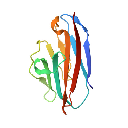
Structure of the N-terminal dimerization domain of CEACAM7.

Bonsor, D.A.Beckett, D.Sundberg, E.J.

(2015) Acta Crystallogr F Struct Biol Commun 71: 1169-1175

  • DOI: https://doi.org/10.1107/S2053230X15013576
  • Primary Citation of Related Structures:  
    4Y89

  • PubMed Abstract: 

    CEACAM7 is a human cellular adhesion protein that is expressed on the surface of colon and rectum epithelial cells and is downregulated in colorectal cancers. It achieves cell adhesion through dimerization of the N-terminal IgV domain. The crystal structure of the N-terminal dimerization domain of CEACAM has been determined at 1.47 Å resolution. The overall fold of CEACAM7 is similar to those of CEACAM1 and CEACAM5; however, there are differences, the most notable of which is an insertion that causes the C'' strand to buckle, leading to the creation of a hydrogen bond in the dimerization interface. The Kdimerization for CEACAM7 determined by sedimentation equilibrium is tenfold tighter than that measured for CEACAM5. These findings suggest that the dimerization affinities of CEACAMs are modulated via sequence variation in the dimerization surface.


  • Organizational Affiliation

    Institute of Human Virology, University of Maryland School of Medicine, Baltimore, MD 21201, USA.


Macromolecules
Find similar proteins by:  (by identity cutoff)  |  3D Structure
Entity ID: 1
MoleculeChains Sequence LengthOrganismDetailsImage
Carcinoembryonic antigen-related cell adhesion molecule 7
A, B, C, D
109Homo sapiensMutation(s): 0 
Gene Names: CEACAM7
UniProt & NIH Common Fund Data Resources
Find proteins for Q14002 (Homo sapiens)
Explore Q14002 
Go to UniProtKB:  Q14002
PHAROS:  Q14002
GTEx:  ENSG00000007306 
Entity Groups  
Sequence Clusters30% Identity50% Identity70% Identity90% Identity95% Identity100% Identity
UniProt GroupQ14002
Sequence Annotations
Expand
  • Reference Sequence
Experimental Data & Validation

Experimental Data

  • Method: X-RAY DIFFRACTION
  • Resolution: 1.47 Å
  • R-Value Free: 0.193 
  • R-Value Work: 0.143 
  • R-Value Observed: 0.145 
  • Space Group: P 1 21 1
Unit Cell:
Length ( Å )Angle ( ˚ )
a = 32.623α = 90
b = 64.886β = 89.99
c = 103.164γ = 90
Software Package:
Software NamePurpose
REFMACrefinement
HKL-2000data reduction
HKL-2000data scaling
PDB_EXTRACTdata extraction
MOLREPphasing

Structure Validation

View Full Validation Report


Entry History 

Deposition Data

Revision History  (Full details and data files)

  • Version 1.0: 2015-09-02
    Type: Initial release
  • Version 1.1: 2015-09-09
    Changes: Database references
  • Version 1.2: 2015-09-16
    Changes: Database references
  • Version 1.3: 2023-09-27
    Changes: Data collection, Database references, Derived calculations, Refinement description