The terminal phycobilisome emitter, LCM: A light-harvesting pigment with a phytochrome chromophore
Tang, K., Ding, W.-L., Hoppner, A., Zhao, C., Zhang, L., Hontani, Y., Kennis, J.T.M., Gartner, W., Scheer, H., Zhou, M., Zhao, K.-H.(2015) Proc Natl Acad Sci U S A 112: 15880-15885
- PubMed: 26669441
- DOI: https://doi.org/10.1073/pnas.1519177113
- Primary Citation of Related Structures:
4XXI, 4XXK - PubMed Abstract:
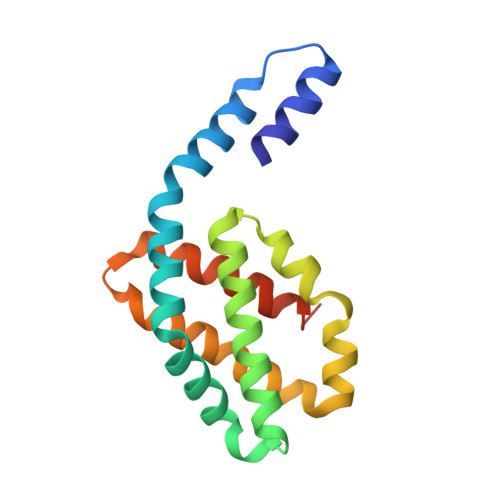
Photosynthesis relies on energy transfer from light-harvesting complexes to reaction centers. Phycobilisomes, the light-harvesting antennas in cyanobacteria and red algae, attach to the membrane via the multidomain core-membrane linker, L(CM). The chromophore domain of L(CM) forms a bottleneck for funneling the harvested energy either productively to reaction centers or, in case of light overload, to quenchers like orange carotenoid protein (OCP) that prevent photodamage. The crystal structure of the solubly modified chromophore domain from Nostoc sp. PCC7120 was resolved at 2.2 Å. Although its protein fold is similar to the protein folds of phycobiliproteins, the phycocyanobilin (PCB) chromophore adopts ZZZssa geometry, which is unknown among phycobiliproteins but characteristic for sensory photoreceptors (phytochromes and cyanobacteriochromes). However, chromophore photoisomerization is inhibited in L(CM) by tight packing. The ZZZssa geometry of the chromophore and π-π stacking with a neighboring Trp account for the functionally relevant extreme spectral red shift of L(CM). Exciton coupling is excluded by the large distance between two PCBs in a homodimer and by preservation of the spectral features in monomers. The structure also indicates a distinct flexibility that could be involved in quenching. The conclusions from the crystal structure are supported by femtosecond transient absorption spectra in solution.
Organizational Affiliation:
State Key Laboratory of Agricultural Microbiology, Huazhong Agricultural University, Wuhan 430070, People's Republic of China; Max Planck Institute for Chemical Energy Conversion, D-45470 Mülheim an der Ruhr, Germany;