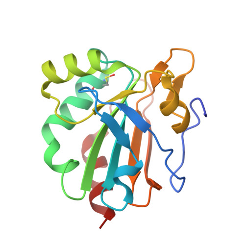
4X1U

The structure of AhpE from Mycobacterium tuberculosis revisited


Experimental Data Snapshot

  • Method: X-RAY DIFFRACTION
  • Resolution: 1.87 Å
  • R-Value Free: 0.183 
  • R-Value Work: 0.151 
  • R-Value Observed: 0.153 

wwPDB Validation   3D Report Full Report


This is version 1.1 of the entry. See complete history

Re-refinement Note

This entry reflects an alternative modeling of the original data in: 1xvw


Literature

Revisiting sulfur H-bonds in proteins: The example of peroxiredoxin AhpE.

van Bergen, L.A.Alonso, M.Pallo, A.Nilsson, L.De Proft, F.Messens, J.

(2016) Sci Rep 6: 30369-30369

  • DOI: https://doi.org/10.1038/srep30369
  • Primary Citation of Related Structures:  
    4X0X, 4X1U

  • PubMed Abstract: 

    In many established methods, identification of hydrogen bonds (H-bonds) is primarily based on pairwise comparison of distances between atoms. These methods often give rise to systematic errors when sulfur is involved. A more accurate method is the non-covalent interaction index, which determines the strength of the H-bonds based on the associated electron density and its gradient. We applied the NCI index on the active site of a single-cysteine peroxiredoxin. We found a different sulfur hydrogen-bonding network to that typically found by established methods, and we propose a more accurate equation for determining sulfur H-bonds based on geometrical criteria. This new algorithm will be implemented in the next release of the widely-used CHARMM program (version 41b), and will be particularly useful for analyzing water molecule-mediated H-bonds involving different atom types. Furthermore, based on the identification of the weakest sulfur-water H-bond, the location of hydrogen peroxide for the nucleophilic attack by the cysteine sulfur can be predicted. In general, current methods to determine H-bonds will need to be reevaluated, thereby leading to better understanding of the catalytic mechanisms in which sulfur chemistry is involved.


  • Organizational Affiliation

    Research Group of General Chemistry, Vrije Universiteit Brussel, 1050 Brussels, Belgium.


Macromolecules
Find similar proteins by:  (by identity cutoff)  |  3D Structure
Entity ID: 1
MoleculeChains Sequence LengthOrganismDetailsImage
Putative peroxiredoxin MT2298
A, B
159Mycobacterium tuberculosisMutation(s): 0 
Gene Names: MT2298
EC: 1.11.1.15
UniProt
Find proteins for P9WIE3 (Mycobacterium tuberculosis (strain ATCC 25618 / H37Rv))
Explore P9WIE3 
Go to UniProtKB:  P9WIE3
Entity Groups  
Sequence Clusters30% Identity50% Identity70% Identity90% Identity95% Identity100% Identity
UniProt GroupP9WIE3
Sequence Annotations
Expand
  • Reference Sequence
Experimental Data & Validation

Experimental Data

  • Method: X-RAY DIFFRACTION
  • Resolution: 1.87 Å
  • R-Value Free: 0.183 
  • R-Value Work: 0.151 
  • R-Value Observed: 0.153 
  • Space Group: I 4
Unit Cell:
Length ( Å )Angle ( ˚ )
a = 147.994α = 90
b = 147.994β = 90
c = 33.711γ = 90
Software Package:
Software NamePurpose
REFMACrefinement
PDB_EXTRACTdata extraction
Cootmodel building
SOLVEphasing
SCALEPACKdata scaling
HKL-2000data reduction

Structure Validation

View Full Validation Report


Entry History & Funding Information

Deposition Data


Funding OrganizationLocationGrant Number
Research Foundation-Flanders (FWO)BelgiumG030512N

Revision History  (Full details and data files)

  • Version 1.0: 2016-07-27
    Type: Initial release
  • Version 1.1: 2016-08-10
    Changes: Database references