The first crystal structure of a glycoside hydrolase family 17 beta-1,3-glucanosyltransferase displays a unique catalytic cleft.
Qin, Z., Yan, Q., Lei, J., Yang, S., Jiang, Z., Wu, S.(2015) Acta Crystallogr D Biol Crystallogr 71: 1714-1724
- PubMed: 26249352
- DOI: https://doi.org/10.1107/S1399004715011037
- Primary Citation of Related Structures:
4WTP, 4WTR, 4WTS - PubMed Abstract:
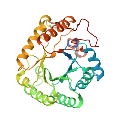
β-1,3-Glucanosyltransferase (EC 2.4.1.-) plays an important role in the formation of branched glucans, as well as in cell-wall assembly and rearrangement in fungi and yeasts. The crystal structures of a novel glycoside hydrolase (GH) family 17 β-1,3-glucanosyltransferase from Rhizomucor miehei (RmBgt17A) and the complexes of its active-site mutant (E189A) with two substrates were solved at resolutions of 1.30, 2.30 and 2.27 Å, respectively. The overall structure of RmBgt17A had the characteristic (β/α)8 TIM-barrel fold. The structures of RmBgt17A and other GH family 17 members were compared: it was found that a conserved subdomain located in the region near helix α6 and part of the catalytic cleft in other GH family 17 members was absent in RmBgt17A. Instead, four amino-acid residues exposed to the surface of the enzyme (Tyr135, Tyr136, Glu158 and His172) were found in the reducing terminus of subsite +2 of RmBgt17A, hindering access to the catalytic cleft. This distinct region of RmBgt17A makes its catalytic cleft shorter than those of other reported GH family 17 enzymes. The complex structures also illustrated that RmBgt17A can only provide subsites -3 to +2. This structural evidence provides a clear explanation of the catalytic mode of RmBgt17A, in which laminaribiose is released from the reducing end of linear β-1,3-glucan and the remaining glucan is transferred to the end of another β-1,3-glucan acceptor. The first crystal structure of a GH family 17 β-1,3-glucanosyltransferase may be useful in studies of the catalytic mechanism of GH family 17 proteins, and provides a basis for further enzymatic engineering or antifungal drug screening.
Organizational Affiliation:
College of Food Science and Nutritional Engineering, Research and Innovation Center of Food Nutrition and Human Health (Beijing), China Agricultural University, Beijing 100083, People's Republic of China.