4WH4

Protein GB1 Quadruple Mutant I6H/N8H/K28H/Q32H


Experimental Data Snapshot

  • Method: X-RAY DIFFRACTION
  • Resolution: 2.20 Å
  • R-Value Free: 0.256 
  • R-Value Work: 0.201 
  • R-Value Observed: 0.207 

wwPDB Validation   3D Report Full Report


This is version 1.3 of the entry. See complete history


Literature

The double-histidine Cu2+-binding motif: a highly rigid, site-specific spin probe for electron spin resonance distance measurements.

Cunningham, T.F.Putterman, M.R.Desai, A.Horne, W.S.Saxena, S.

(2015) Angew Chem Int Ed Engl 54: 6330-6334

  • DOI: https://doi.org/10.1002/anie.201501968
  • Primary Citation of Related Structures:  
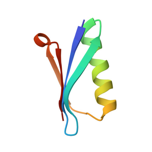
    4WH4

  • PubMed Abstract: 

    The development of ESR methods that measure long-range distance distributions has advanced biophysical research. However, the spin labels commonly employed are highly flexible, which leads to ambiguity in relating ESR measurements to protein-backbone structure. Herein we present the double-histidine (dHis) Cu(2+)-binding motif as a rigid spin probe for double electron-electron resonance (DEER) distance measurements. The spin label is assembled in situ from natural amino acid residues and a metal salt, requires no postexpression synthetic modification, and provides distance distributions that are dramatically narrower than those found with the commonly used protein spin label. Simple molecular modeling based on an X-ray crystal structure of an unlabeled protein led to a predicted most probable distance within 0.5 Å of the experimental value. Cu(2+) DEER with the dHis motif shows great promise for the resolution of precise, unambiguous distance constraints that relate directly to protein-backbone structure and flexibility.


  • Organizational Affiliation

    Department of Chemistry, University of Pittsburgh, 219 Parkman Avenue, Pittsburgh, PA 15260 (USA).


Macromolecules
Find similar proteins by:  (by identity cutoff)  |  3D Structure
Entity ID: 1
MoleculeChains Sequence LengthOrganismDetailsImage
Immunoglobulin G-binding protein G
A, B
56Streptococcus sp. 'group GMutation(s): 4 
Gene Names: spg
UniProt
Find proteins for P19909 (Streptococcus sp. group G)
Explore P19909 
Go to UniProtKB:  P19909
Entity Groups  
Sequence Clusters30% Identity50% Identity70% Identity90% Identity95% Identity100% Identity
UniProt GroupP19909
Sequence Annotations
Expand
  • Reference Sequence
Experimental Data & Validation

Experimental Data

  • Method: X-RAY DIFFRACTION
  • Resolution: 2.20 Å
  • R-Value Free: 0.256 
  • R-Value Work: 0.201 
  • R-Value Observed: 0.207 
  • Space Group: P 31 2 1
Unit Cell:
Length ( Å )Angle ( ˚ )
a = 74.287α = 90
b = 74.287β = 90
c = 41.32γ = 120
Software Package:
Software NamePurpose
PHENIXrefinement

Structure Validation

View Full Validation Report


Entry History & Funding Information

Deposition Data


Funding OrganizationLocationGrant Number
National Science Foundation (NSF, United States)United StatesMCB 1157712

Revision History  (Full details and data files)

  • Version 1.0: 2015-08-05
    Type: Initial release
  • Version 1.1: 2017-09-06
    Changes: Author supporting evidence, Database references, Derived calculations
  • Version 1.2: 2019-11-27
    Changes: Author supporting evidence
  • Version 1.3: 2023-09-27
    Changes: Data collection, Database references, Refinement description