4WFH

Human TRAAK K+ channel in a Tl+ bound nonconductive conformation


Experimental Data Snapshot

  • Method: X-RAY DIFFRACTION
  • Resolution: 3.01 Å
  • R-Value Free: 0.238 
  • R-Value Work: 0.203 
  • R-Value Observed: 0.205 

wwPDB Validation   3D Report Full Report


This is version 1.3 of the entry. See complete history


Literature

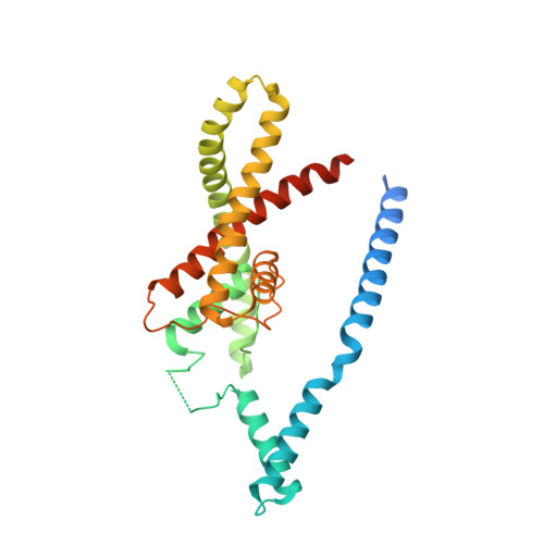
Physical mechanism for gating and mechanosensitivity of the human TRAAK K+ channel.

Brohawn, S.G.Campbell, E.B.MacKinnon, R.

(2014) Nature 516: 126-130

  • DOI: https://doi.org/10.1038/nature14013

  • PubMed Abstract: 

    Activation of mechanosensitive ion channels by physical force underlies many physiological processes including the sensation of touch, hearing and pain. TRAAK (also known as KCNK4) ion channels are neuronally expressed members of the two-pore domain K(+) (K2P) channel family and are mechanosensitive. They are involved in controlling mechanical and temperature nociception in mice. Mechanosensitivity of TRAAK is mediated directly through the lipid bilayer--it is a membrane-tension-gated channel. However, the molecular mechanism of TRAAK channel gating and mechanosensitivity is unknown. Here we present crystal structures of TRAAK in conductive and non-conductive conformations defined by the presence of permeant ions along the conduction pathway. In the non-conductive state, a lipid acyl chain accesses the channel cavity through a 5 Å-wide lateral opening in the membrane inner leaflet and physically blocks ion passage. In the conductive state, rotation of a transmembrane helix (TM4) about a central hinge seals the intramembrane opening, preventing lipid block of the cavity and permitting ion entry. Additional rotation of a membrane interacting TM2-TM3 segment, unique to mechanosensitive K2Ps, against TM4 may further stabilize the conductive conformation. Comparison of the structures reveals a biophysical explanation for TRAAK mechanosensitivity--an expansion in cross-sectional area up to 2.7 nm(2) in the conductive state is expected to create a membrane-tension-dependent energy difference between conformations that promotes force activation. Our results show how tension of the lipid bilayer can be harnessed to control gating and mechanosensitivity of a eukaryotic ion channel.


  • Organizational Affiliation

    Laboratory of Molecular Neurobiology and Biophysics and Howard Hughes Medical Institute, The Rockefeller University, 1230 York Avenue, New York, New York 10065, USA.


Macromolecules
Find similar proteins by:  (by identity cutoff)  |  3D Structure
Entity ID: 1
MoleculeChains Sequence LengthOrganismDetailsImage
Potassium channel subfamily K member 4
A, B
299Homo sapiensMutation(s): 2 
Gene Names: KCNK4TRAAK
Membrane Entity: Yes 
UniProt & NIH Common Fund Data Resources
Find proteins for Q9NYG8 (Homo sapiens)
Explore Q9NYG8 
Go to UniProtKB:  Q9NYG8
PHAROS:  Q9NYG8
GTEx:  ENSG00000182450 
Entity Groups  
Sequence Clusters30% Identity50% Identity70% Identity90% Identity95% Identity100% Identity
UniProt GroupQ9NYG8
Sequence Annotations
Expand
  • Reference Sequence
Find similar proteins by:  (by identity cutoff)  |  3D Structure
Entity ID: 2
MoleculeChains Sequence LengthOrganismDetailsImage
ANTI-TRAAK ANTIBODY 13E9 FAB FRAGMENT LIGHT CHAINC [auth D],
E [auth F]
211Mus musculusMutation(s): 0 
Entity Groups  
Sequence Clusters30% Identity50% Identity70% Identity90% Identity95% Identity100% Identity
Sequence Annotations
Expand
  • Reference Sequence
Find similar proteins by:  (by identity cutoff)  |  3D Structure
Entity ID: 3
MoleculeChains Sequence LengthOrganismDetailsImage
ANTI-TRAAK ANTIBODY 13E9 FAB FRAGMENT HEAVY CHAIND [auth E],
F [auth G]
217Mus musculusMutation(s): 0 
Entity Groups  
Sequence Clusters30% Identity50% Identity70% Identity90% Identity95% Identity100% Identity
Sequence Annotations
Expand
  • Reference Sequence
Small Molecules
Ligands 3 Unique
IDChains Name / Formula / InChI Key2D Diagram3D Interactions
TL
Query on TL

Download Ideal Coordinates CCD File 
G [auth A]
H [auth A]
I [auth A]
J [auth A]
M [auth A]
G [auth A],
H [auth A],
I [auth A],
J [auth A],
M [auth A],
N [auth B],
O [auth B]
THALLIUM (I) ION
Tl
ZLUSCZLCHQSJRU-UHFFFAOYSA-N
D10
Query on D10

Download Ideal Coordinates CCD File 
P [auth B]DECANE
C10 H22
DIOQZVSQGTUSAI-UHFFFAOYSA-N
CA
Query on CA

Download Ideal Coordinates CCD File 
K [auth A],
L [auth A],
Q [auth G]
CALCIUM ION
Ca
BHPQYMZQTOCNFJ-UHFFFAOYSA-N
Experimental Data & Validation

Experimental Data

  • Method: X-RAY DIFFRACTION
  • Resolution: 3.01 Å
  • R-Value Free: 0.238 
  • R-Value Work: 0.203 
  • R-Value Observed: 0.205 
  • Space Group: P 1 21 1
Unit Cell:
Length ( Å )Angle ( ˚ )
a = 80.972α = 90
b = 138.755β = 94.64
c = 96.759γ = 90
Software Package:
Software NamePurpose
REFMACrefinement

Structure Validation

View Full Validation Report


Entry History 

Deposition Data

Revision History  (Full details and data files)

  • Version 1.0: 2014-12-03
    Type: Initial release
  • Version 1.1: 2014-12-17
    Changes: Database references
  • Version 1.2: 2015-02-04
    Changes: Derived calculations
  • Version 1.3: 2023-12-27
    Changes: Data collection, Database references, Derived calculations, Other, Refinement description, Source and taxonomy, Structure summary