4W8N

The crystal structure of hemagglutinin from a swine influenza virus (A/swine/Missouri/2124514/2006)


Experimental Data Snapshot

  • Method: X-RAY DIFFRACTION
  • Resolution: 2.90 Å
  • R-Value Free: 0.262 
  • R-Value Work: 0.233 
  • R-Value Observed: 0.234 

wwPDB Validation   3D Report Full Report


Ligand Structure Quality Assessment 


This is version 2.1 of the entry. See complete history


Literature

Assessment of transmission, pathogenesis and adaptation of H2 subtype influenza viruses in ferrets.

Pappas, C.Yang, H.Carney, P.J.Pearce, M.B.Katz, J.M.Stevens, J.Tumpey, T.M.

(2015) Virology 477C: 61-71

  • DOI: https://doi.org/10.1016/j.virol.2015.01.002
  • Primary Citation of Related Structures:  
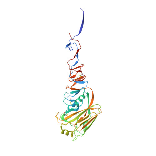
    4W8N

  • PubMed Abstract: 

    After their disappearance from the human population in 1968, influenza H2 viruses have continued to circulate in the natural avian reservoir. The isolation of this virus subtype from multiple bird species as well as swine highlights the need to better understand the potential of these viruses to spread and cause disease in humans. Here we analyzed the virulence, transmissibility and receptor-binding preference of two avian influenza H2 viruses (H2N2 and H2N3) and compared them to a swine H2N3 (A/swine/Missouri/2124514/2006 [swMO]), and a human H2N2 (A/England/10/1967 [Eng/67]) virus using the ferret model as a mammalian host. Both avian H2 viruses possessed the capacity to spread efficiently between cohoused ferrets, and the swine (swMO) and human (Eng/67) viruses transmitted to naïve ferrets by respiratory droplets. Further characterization of the swMO hemagglutinin (HA) by x-ray crystallography and glycan microarray array identified receptor-specific adaptive mutations. As influenza virus quasispecies dynamics during transmission have not been well characterized, we sequenced nasal washes collected during transmission studies to better understand experimental adaptation of H2 HA. The avian H2 viruses isolated from ferret nasal washes contained mutations in the HA1, including a Gln226Leu substitution, which is a mutation associated with α2,6 sialic acid (human-like) binding preference. These results suggest that the molecular structure of HA in viruses of the H2 subtype continue to have the potential to adapt to a mammalian host and become transmissible, after acquiring additional genetic markers.


  • Organizational Affiliation

    Influenza Division, NCIRD, Centers for Disease Control and Prevention, 1600 Clifton Road NE, Atlanta, GA 30333, USA.


Macromolecules
Find similar proteins by:  (by identity cutoff)  |  3D Structure
Entity ID: 1
MoleculeChains Sequence LengthOrganismDetailsImage
Hemagglutinin
A, C, E
330Influenza A virus (A/swine/Missouri/2124514/2006(H2N3))Mutation(s): 0 
Gene Names: HA
UniProt
Find proteins for A9YN66 (Influenza A virus)
Explore A9YN66 
Go to UniProtKB:  A9YN66
Entity Groups  
Sequence Clusters30% Identity50% Identity70% Identity90% Identity95% Identity100% Identity
UniProt GroupA9YN66
Sequence Annotations
Expand
  • Reference Sequence
Find similar proteins by:  (by identity cutoff)  |  3D Structure
Entity ID: 2
MoleculeChains Sequence LengthOrganismDetailsImage
Hemagglutinin
B, D, F
179Influenza A virus (A/swine/Missouri/2124514/2006(H2N3))Mutation(s): 0 
Gene Names: HA
UniProt
Find proteins for A9YN66 (Influenza A virus)
Explore A9YN66 
Go to UniProtKB:  A9YN66
Entity Groups  
Sequence Clusters30% Identity50% Identity70% Identity90% Identity95% Identity100% Identity
UniProt GroupA9YN66
Sequence Annotations
Expand
  • Reference Sequence
Small Molecules
Ligands 1 Unique
IDChains Name / Formula / InChI Key2D Diagram3D Interactions
NAG
Query on NAG

Download Ideal Coordinates CCD File 
G [auth A]
H [auth A]
I [auth A]
J [auth B]
K [auth C]
G [auth A],
H [auth A],
I [auth A],
J [auth B],
K [auth C],
L [auth C],
M [auth C],
N [auth C],
O [auth E],
P [auth E],
Q [auth E]
2-acetamido-2-deoxy-beta-D-glucopyranose
C8 H15 N O6
OVRNDRQMDRJTHS-FMDGEEDCSA-N
Experimental Data & Validation

Experimental Data

  • Method: X-RAY DIFFRACTION
  • Resolution: 2.90 Å
  • R-Value Free: 0.262 
  • R-Value Work: 0.233 
  • R-Value Observed: 0.234 
  • Space Group: P 21 21 21
Unit Cell:
Length ( Å )Angle ( ˚ )
a = 70.291α = 90
b = 160.028β = 90
c = 170.602γ = 90
Software Package:
Software NamePurpose
REFMACrefinement

Structure Validation

View Full Validation Report


Ligand Structure Quality Assessment 


Entry History 

Deposition Data

Revision History  (Full details and data files)

  • Version 1.0: 2015-02-11
    Type: Initial release
  • Version 1.1: 2015-03-04
    Changes: Database references
  • Version 2.0: 2020-07-29
    Type: Remediation
    Reason: Carbohydrate remediation
    Changes: Advisory, Atomic model, Data collection, Database references, Derived calculations, Non-polymer description, Other, Refinement description, Source and taxonomy, Structure summary
  • Version 2.1: 2023-12-27
    Changes: Advisory, Data collection, Database references, Structure summary