4UTS

Room temperature crystal structure of the fast switching M159T mutant of fluorescent protein Dronpa


Experimental Data Snapshot

  • Method: X-RAY DIFFRACTION
  • Resolution: 2.03 Å
  • R-Value Free: 0.213 
  • R-Value Work: 0.172 
  • R-Value Observed: 0.174 

wwPDB Validation   3D Report Full Report


This is version 2.1 of the entry. See complete history


Literature

Room Temperature Crystal Structure of the Fast Switching M159T Mutant of the Fluorescent Protein Dronpa.

Kaucikas, M.Fitzpatrick, A.Bryan, E.Struve, A.Henning, R.Kosheleva, I.Srajer, V.Groenhof, G.Van Thor, J.J.

(2015) Proteins 83: 397

  • DOI: https://doi.org/10.1002/prot.24742
  • Primary Citation of Related Structures:  
    4UTS

  • PubMed Abstract: 

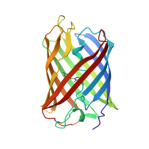
    The fluorescent protein Dronpa undergoes reversible photoswitching reactions between the bright "on" and dark "off" states via photoisomerization and proton transfer reactions. We report the room temperature crystal structure of the fast switching Met159Thr mutant of Dronpa at 2.0-Å resolution in the bright on state. Structural differences with the wild type include shifted backbone positions of strand β8 containing Thr159 as well as an altered A-C dimer interface involving strands β7, β8, β10, and β11. The Met159Thr mutation increases the cavity volume for the p-hydroxybenzylidene-imidazolinone chromophore as a result of both the side chain difference and the backbone positional differences.


  • Organizational Affiliation

    Division of Molecular Biosciences, Imperial College London, London, SW7 2AZ, United Kingdom.


Macromolecules
Find similar proteins by:  (by identity cutoff)  |  3D Structure
Entity ID: 1
MoleculeChains Sequence LengthOrganismDetailsImage
FLUORESCENT PROTEIN DRONPA
A, B, C, D
214Echinophyllia sp. SC22Mutation(s): 2 
UniProt
Find proteins for Q5TLG6 (Echinophyllia sp. SC22)
Explore Q5TLG6 
Go to UniProtKB:  Q5TLG6
Entity Groups  
Sequence Clusters30% Identity50% Identity70% Identity90% Identity95% Identity100% Identity
UniProt GroupQ5TLG6
Sequence Annotations
Expand
  • Reference Sequence
Small Molecules
Modified Residues  1 Unique
IDChains TypeFormula2D DiagramParent
GYC
Query on GYC
A, B, C, D
L-PEPTIDE LINKINGC14 H15 N3 O4 SCYS, TYR, GLY
Experimental Data & Validation

Experimental Data

  • Method: X-RAY DIFFRACTION
  • Resolution: 2.03 Å
  • R-Value Free: 0.213 
  • R-Value Work: 0.172 
  • R-Value Observed: 0.174 
  • Space Group: P 21 21 21
Unit Cell:
Length ( Å )Angle ( ˚ )
a = 75.655α = 90
b = 111.14β = 90
c = 117.572γ = 90
Software Package:
Software NamePurpose
REFMACrefinement
MOSFLMdata reduction
SCALAdata scaling
MOLREPphasing

Structure Validation

View Full Validation Report


Entry History 

Deposition Data

Revision History  (Full details and data files)

  • Version 1.0: 2015-06-03
    Type: Initial release
  • Version 1.1: 2019-10-23
    Changes: Data collection, Database references, Derived calculations, Other
  • Version 2.0: 2023-11-15
    Changes: Advisory, Atomic model, Data collection, Database references, Derived calculations
  • Version 2.1: 2024-01-10
    Changes: Refinement description