4U61

Trichodysplasia spinulosa-associated polyomavirus (TSPyV) VP1 in complex with 6'-sialyllactose


Experimental Data Snapshot

  • Method: X-RAY DIFFRACTION
  • Resolution: 1.65 Å
  • R-Value Free: 0.184 
  • R-Value Work: 0.165 
  • R-Value Observed: 0.166 

wwPDB Validation   3D Report Full Report


Ligand Structure Quality Assessment 


This is version 2.1 of the entry. See complete history


Literature

Trichodysplasia spinulosa-Associated Polyomavirus Uses a Displaced Binding Site on VP1 to Engage Sialylated Glycolipids.

Stroh, L.J.Gee, G.V.Blaum, B.S.Dugan, A.S.Feltkamp, M.C.Atwood, W.J.Stehle, T.

(2015) PLoS Pathog 11: e1005112-e1005112

  • DOI: https://doi.org/10.1371/journal.ppat.1005112
  • Primary Citation of Related Structures:  
    4U5Z, 4U60, 4U61, 4U62

  • PubMed Abstract: 

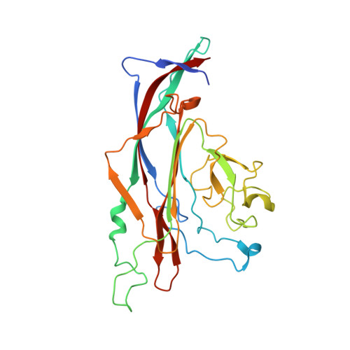
    Trichodysplasia spinulosa-associated Polyomavirus (TSPyV) was isolated from a patient suffering from trichodysplasia spinulosa, a skin disease that can appear in severely immunocompromised patients. While TSPyV is one of the five members of the polyomavirus family that are directly linked to a human disease, details about molecular recognition events, the viral entry pathway, and intracellular trafficking events during TSPyV infection remain unknown. Here we have used a structure-function approach to shed light on the first steps of TSPyV infection. We established by cell binding and pseudovirus infection studies that TSPyV interacts with sialic acids during attachment and/or entry. Subsequently, we solved high-resolution X-ray structures of the major capsid protein VP1 of TSPyV in complex with three different glycans, the branched GM1 glycan, and the linear trisaccharides α2,3- and α2,6-sialyllactose. The terminal sialic acid of all three glycans is engaged in a unique binding site on TSPyV VP1, which is positioned about 18 Å from established sialic acid binding sites of other polyomaviruses. Structure-based mutagenesis of sialic acid-binding residues leads to reduction in cell attachment and pseudovirus infection, demonstrating the physiological relevance of the TSPyV VP1-glycan interaction. Furthermore, treatments of cells with inhibitors of N-, O-linked glycosylation, and glycosphingolipid synthesis suggest that glycolipids play an important role during TSPyV infection. Our findings elucidate the first molecular recognition events of cellular infection with TSPyV and demonstrate that receptor recognition by polyomaviruses is highly variable not only in interactions with sialic acid itself, but also in the location of the binding site.


  • Organizational Affiliation

    Interfaculty Institute of Biochemistry, University of Tübingen, Tübingen, Germany.


Macromolecules
Find similar proteins by:  (by identity cutoff)  |  3D Structure
Entity ID: 1
MoleculeChains Sequence LengthOrganismDetailsImage
Structural protein VP1
A, B, C, D, E
A, B, C, D, E, F, G, H, I, J
280Trichodysplasia spinulosa-associated polyomavirusMutation(s): 0 
Gene Names: VP1
UniProt
Find proteins for E2ESL7 (Trichodysplasia spinulosa-associated polyomavirus)
Explore E2ESL7 
Go to UniProtKB:  E2ESL7
Entity Groups  
Sequence Clusters30% Identity50% Identity70% Identity90% Identity95% Identity100% Identity
UniProt GroupE2ESL7
Sequence Annotations
Expand
  • Reference Sequence
Oligosaccharides

Help

Entity ID: 2
MoleculeChains Length2D Diagram Glycosylation3D Interactions
N-acetyl-alpha-neuraminic acid-(2-6)-beta-D-galactopyranose
K
2N/A
Glycosylation Resources
GlyTouCan:  G63069TR
GlyCosmos:  G63069TR
GlyGen:  G63069TR
Small Molecules
Ligands 2 Unique
IDChains Name / Formula / InChI Key2D Diagram3D Interactions
SIA
Query on SIA

Download Ideal Coordinates CCD File 
L [auth B]
N [auth C]
O [auth E]
S [auth H]
T [auth I]
L [auth B],
N [auth C],
O [auth E],
S [auth H],
T [auth I],
U [auth J]
N-acetyl-alpha-neuraminic acid
C11 H19 N O9
SQVRNKJHWKZAKO-YRMXFSIDSA-N
GOL
Query on GOL

Download Ideal Coordinates CCD File 
M [auth C],
P [auth E],
Q [auth E],
R [auth H]
GLYCEROL
C3 H8 O3
PEDCQBHIVMGVHV-UHFFFAOYSA-N
Experimental Data & Validation

Experimental Data

  • Method: X-RAY DIFFRACTION
  • Resolution: 1.65 Å
  • R-Value Free: 0.184 
  • R-Value Work: 0.165 
  • R-Value Observed: 0.166 
  • Space Group: P 21 21 21
Unit Cell:
Length ( Å )Angle ( ˚ )
a = 139.56α = 90
b = 146.35β = 90
c = 151.54γ = 90
Software Package:
Software NamePurpose
REFMACrefinement
Cootmodel building
XDSdata reduction
PHASERphasing
XSCALEdata scaling

Structure Validation

View Full Validation Report


Ligand Structure Quality Assessment 


Entry History 

Deposition Data

Revision History  (Full details and data files)

  • Version 1.0: 2015-08-05
    Type: Initial release
  • Version 1.1: 2015-09-02
    Changes: Database references
  • Version 2.0: 2020-07-29
    Type: Remediation
    Reason: Carbohydrate remediation
    Changes: Atomic model, Data collection, Derived calculations, Structure summary
  • Version 2.1: 2023-12-20
    Changes: Data collection, Database references, Derived calculations, Refinement description, Structure summary