4U4G

Structure of GluA2* in complex with competitive antagonist ZK 200775


Experimental Data Snapshot

  • Method: X-RAY DIFFRACTION
  • Resolution: 4.49 Å
  • R-Value Free: 0.248 
  • R-Value Work: 0.217 
  • R-Value Observed: 0.218 

wwPDB Validation   3D Report Full Report


This is version 2.1 of the entry. See complete history


Literature

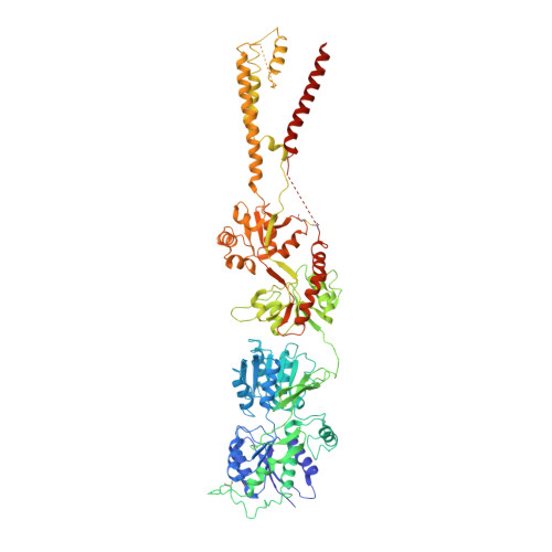
Structure of an agonist-bound ionotropic glutamate receptor.

Yelshanskaya, M.V.Li, M.Sobolevsky, A.I.

(2014) Science 345: 1070-1074

  • DOI: https://doi.org/10.1126/science.1256508
  • Primary Citation of Related Structures:  
    4U4F, 4U4G

  • PubMed Abstract: 

    Ionotropic glutamate receptors (iGluRs) mediate most excitatory neurotransmission in the central nervous system and function by opening their ion channel in response to binding of agonist glutamate. Here, we report a structure of a homotetrameric rat GluA2 receptor in complex with partial agonist (S)-5-nitrowillardiine. Comparison of this structure with the closed-state structure in complex with competitive antagonist ZK 200775 suggests conformational changes that occur during iGluR gating. Guided by the structures, we engineered disulfide cross-links to probe domain interactions that are important for iGluR gating events. The combination of structural information, kinetic modeling, and biochemical and electrophysiological experiments provides insight into the mechanism of iGluR gating.


  • Organizational Affiliation

    Department of Biochemistry and Molecular Biophysics, Columbia University, 650 West 168th Street, New York, NY 10032, USA.


Macromolecules
Find similar proteins by:  (by identity cutoff)  |  3D Structure
Entity ID: 1
MoleculeChains Sequence LengthOrganismDetailsImage
Glutamate receptor 2
A, B, C, D
822Rattus norvegicusMutation(s): 0 
Gene Names: Gria2Glur2
Membrane Entity: Yes 
UniProt
Find proteins for P19491 (Rattus norvegicus)
Explore P19491 
Go to UniProtKB:  P19491
Entity Groups  
Sequence Clusters30% Identity50% Identity70% Identity90% Identity95% Identity100% Identity
UniProt GroupP19491
Sequence Annotations
Expand
  • Reference Sequence
Oligosaccharides

Help

Entity ID: 2
MoleculeChains Length2D Diagram Glycosylation3D Interactions
beta-D-mannopyranose-(1-3)-beta-D-mannopyranose-(1-4)-2-acetamido-2-deoxy-beta-D-glucopyranose-(1-4)-2-acetamido-2-deoxy-beta-D-glucopyranose
E, F, G, H
4N-Glycosylation
Glycosylation Resources
GlyTouCan:  G31886NL
GlyCosmos:  G31886NL
GlyGen:  G31886NL
Experimental Data & Validation

Experimental Data

  • Method: X-RAY DIFFRACTION
  • Resolution: 4.49 Å
  • R-Value Free: 0.248 
  • R-Value Work: 0.217 
  • R-Value Observed: 0.218 
  • Space Group: P 1 21 1
Unit Cell:
Length ( Å )Angle ( ˚ )
a = 91.999α = 90
b = 310.36β = 94.82
c = 109.502γ = 90
Software Package:
Software NamePurpose
PHENIXrefinement
PHASERphasing
HKL-2000data reduction
HKL-2000data scaling

Structure Validation

View Full Validation Report


Entry History & Funding Information

Deposition Data


Funding OrganizationLocationGrant Number
National Institutes of Health/National Institute of Neurological Disorders and Stroke (NIH/NINDS)United StatesNS083660

Revision History  (Full details and data files)

  • Version 1.0: 2014-10-22
    Type: Initial release
  • Version 1.1: 2015-02-11
    Changes: Advisory
  • Version 1.2: 2016-12-21
    Changes: Structure summary
  • Version 1.3: 2017-09-13
    Changes: Author supporting evidence
  • Version 1.4: 2017-11-22
    Changes: Refinement description
  • Version 1.5: 2019-12-18
    Changes: Author supporting evidence
  • Version 2.0: 2020-07-29
    Type: Remediation
    Reason: Carbohydrate remediation
    Changes: Advisory, Atomic model, Data collection, Derived calculations, Structure summary
  • Version 2.1: 2023-09-27
    Changes: Data collection, Database references, Derived calculations, Refinement description, Structure summary