4TW2

Crystal Structure of SCARB2 in Neural Condition (pH7.5)


Experimental Data Snapshot

  • Method: X-RAY DIFFRACTION
  • Resolution: 2.89 Å
  • R-Value Free: 0.293 
  • R-Value Work: 0.244 
  • R-Value Observed: 0.246 

wwPDB Validation   3D Report Full Report


Ligand Structure Quality Assessment 


This is version 3.0 of the entry. See complete history


Literature

Molecular mechanism of SCARB2-mediated attachment and uncoating of EV71

Dang, M.Wang, X.Wang, Q.Wang, Y.Lin, J.Sun, Y.Li, X.Zhang, L.Lou, Z.Wang, J.Rao, Z.

(2014) Protein Cell 5: 692-703

  • DOI: https://doi.org/10.1007/s13238-014-0087-3
  • Primary Citation of Related Structures:  
    4TVZ, 4TW0, 4TW2

  • PubMed Abstract: 

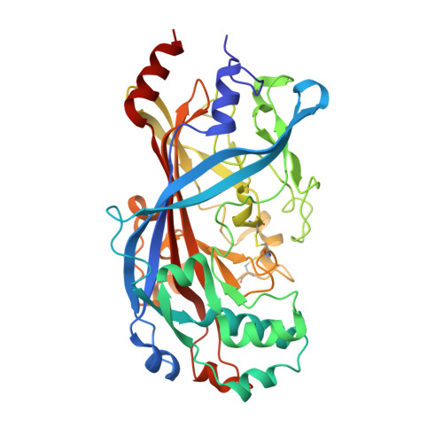
    Unlike the well-established picture for the entry of enveloped viruses, the mechanism of cellular entry of non-enveloped eukaryotic viruses remains largely mysterious. Picornaviruses are representative models for such viruses, and initiate this entry process by their functional receptors. Here we present the structural and functional studies of SCARB2, a functional receptor of the important human enterovirus 71 (EV71). SCARB2 is responsible for attachment as well as uncoating of EV71. Differences in the structures of SCARB2 under neutral and acidic conditions reveal that SCARB2 undergoes a pivotal pH-dependent conformational change which opens a lipid-transfer tunnel to mediate the expulsion of a hydrophobic pocket factor from the virion, a pre-requisite for uncoating. We have also identified the key residues essential for attachment to SCARB2, identifying the canyon region of EV71 as mediating the receptor interaction. Together these results provide a clear understanding of cellular attachment and initiation of uncoating for enteroviruses.


  • Organizational Affiliation

    National Laboratory of Macromolecules, Institute of Biophysics, Chinese Academy of Science, Beijing, 100101, China.


Macromolecules
Find similar proteins by:  (by identity cutoff)  |  3D Structure
Entity ID: 1
MoleculeChains Sequence LengthOrganismDetailsImage
Scavenger receptor class B member 2
A, B
394Homo sapiensMutation(s): 0 
Gene Names: SCARB2CD36L2LIMP2LIMPII
UniProt & NIH Common Fund Data Resources
Find proteins for Q14108 (Homo sapiens)
Explore Q14108 
Go to UniProtKB:  Q14108
PHAROS:  Q14108
GTEx:  ENSG00000138760 
Entity Groups  
Sequence Clusters30% Identity50% Identity70% Identity90% Identity95% Identity100% Identity
UniProt GroupQ14108
Sequence Annotations
Expand
  • Reference Sequence
Oligosaccharides

Help

Entity ID: 2
MoleculeChains Length2D Diagram Glycosylation3D Interactions
alpha-D-mannopyranose-(1-3)-[alpha-D-mannopyranose-(1-6)]beta-D-mannopyranose-(1-4)-2-acetamido-2-deoxy-beta-D-glucopyranose-(1-4)-2-acetamido-2-deoxy-beta-D-glucopyranose
C
5N-Glycosylation
Glycosylation Resources
GlyTouCan:  G22768VO
GlyCosmos:  G22768VO
GlyGen:  G22768VO
Entity ID: 3
MoleculeChains Length2D Diagram Glycosylation3D Interactions
2-acetamido-2-deoxy-beta-D-glucopyranose-(1-4)-2-acetamido-2-deoxy-beta-D-glucopyranose
D, F, J, K
2N-Glycosylation
Glycosylation Resources
GlyTouCan:  G42666HT
GlyCosmos:  G42666HT
GlyGen:  G42666HT
Entity ID: 4
MoleculeChains Length2D Diagram Glycosylation3D Interactions
beta-D-mannopyranose-(1-4)-2-acetamido-2-deoxy-beta-D-glucopyranose-(1-4)-2-acetamido-2-deoxy-beta-D-glucopyranose
E
3N-Glycosylation
Glycosylation Resources
GlyTouCan:  G15407YE
GlyCosmos:  G15407YE
GlyGen:  G15407YE
Entity ID: 5
MoleculeChains Length2D Diagram Glycosylation3D Interactions
alpha-D-mannopyranose-(1-6)-beta-D-mannopyranose-(1-4)-2-acetamido-2-deoxy-beta-D-glucopyranose-(1-4)-2-acetamido-2-deoxy-beta-D-glucopyranose
G
4N-Glycosylation
Glycosylation Resources
GlyTouCan:  G22573RC
GlyCosmos:  G22573RC
GlyGen:  G22573RC
Entity ID: 6
MoleculeChains Length2D Diagram Glycosylation3D Interactions
alpha-D-mannopyranose-(1-3)-beta-D-mannopyranose-(1-4)-2-acetamido-2-deoxy-beta-D-glucopyranose-(1-4)-2-acetamido-2-deoxy-beta-D-glucopyranose
H, I, L
4N-Glycosylation
Glycosylation Resources
GlyTouCan:  G81315DD
GlyCosmos:  G81315DD
GlyGen:  G81315DD
Experimental Data & Validation

Experimental Data

  • Method: X-RAY DIFFRACTION
  • Resolution: 2.89 Å
  • R-Value Free: 0.293 
  • R-Value Work: 0.244 
  • R-Value Observed: 0.246 
  • Space Group: P 1 21 1
Unit Cell:
Length ( Å )Angle ( ˚ )
a = 57.796α = 90
b = 100.317β = 98.69
c = 105.173γ = 90
Software Package:
Software NamePurpose
PHENIXrefinement
SCALEPACKdata reduction
Cootmodel building
PDB_EXTRACTdata extraction
PHASERphasing
SCALEPACKdata scaling

Structure Validation

View Full Validation Report


Ligand Structure Quality Assessment 


Entry History & Funding Information

Deposition Data


Funding OrganizationLocationGrant Number
Institute of Biophysics, Chinese Academy of ScienceChina2014CB542800

Revision History  (Full details and data files)

  • Version 1.0: 2015-07-08
    Type: Initial release
  • Version 1.1: 2015-07-15
    Changes: Derived calculations
  • Version 1.2: 2020-01-29
    Changes: Data collection, Derived calculations, Refinement description, Source and taxonomy
  • Version 2.0: 2020-07-29
    Type: Remediation
    Reason: Carbohydrate remediation
    Changes: Atomic model, Data collection, Derived calculations, Structure summary
  • Version 3.0: 2023-11-08
    Changes: Atomic model, Data collection, Database references, Derived calculations, Refinement description, Structure summary